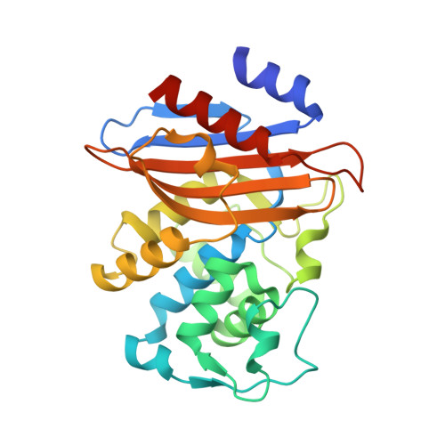
2WK0

Crystal structure of the class A beta-lactamase BS3 inhibited by 6- beta-iodopenicillanate.


Experimental Data Snapshot

  • Method: X-RAY DIFFRACTION
  • Resolution: 1.65 Å
  • R-Value Free: 0.211 
  • R-Value Work: 0.189 
  • R-Value Observed: 0.190 

Starting Model: experimental
View more details

wwPDB Validation   3D Report Full Report


Ligand Structure Quality Assessment 


This is version 1.3 of the entry. See complete history


Literature

Structural Basis of the Inhibition of Class a Beta-Lactamases and Penicillin-Binding Proteins by 6-Beta-Iodopenicillanate.

Sauvage, E.Zervosen, A.Dive, G.Herman, R.Amoroso, A.Joris, B.Fonze, E.Pratt, R.F.Luxen, A.Charlier, P.Kerff, F.

(2009) J Am Chem Soc 131: 15262

  • DOI: https://doi.org/10.1021/ja9051526
  • Primary Citation of Related Structures:  
    2WK0, 2WKE

  • PubMed Abstract: 

    6-Beta-halogenopenicillanates are powerful, irreversible inhibitors of various beta-lactamases and penicillin-binding proteins. Upon acylation of these enzymes, the inhibitors are thought to undergo a structural rearrangement associated with the departure of the iodide and formation of a dihydrothiazine ring, but, to date, no structural evidence has proven this. 6-Beta-iodopenicillanic acid (BIP) is shown here to be an active antibiotic against various bacterial strains and an effective inhibitor of the class A beta-lactamase of Bacillus subtilis BS3 (BS3) and the D,D-peptidase of Actinomadura R39 (R39). Crystals of BS3 and of R39 were soaked with a solution of BIP and their structures solved at 1.65 and 2.2 A, respectively. The beta-lactam and the thiazolidine rings of BIP are indeed found to be fused into a dihydrothiazine ring that can adopt two stable conformations at these active sites. The rearranged BIP is observed in one conformation in the BS3 active site and in two monomers of the asymmetric unit of R39, and is observed in the other conformation in the other two monomers of the asymmetric unit of R39. The BS3 structure reveals a new mode of carboxylate interaction with a class A beta-lactamase active site that should be of interest in future inhibitor design.


  • Organizational Affiliation

    Centre d'Ingénierie des Protéines and Centre de Recherches du Cyclotron, Université de Liège, B-4000 Sart Tilman, Belgium. [email protected]


Macromolecules
Find similar proteins by:  (by identity cutoff)  |  3D Structure
Entity ID: 1
MoleculeChains Sequence LengthOrganismDetailsImage
BETA-LACTAMASE
A, B
265Bacillus licheniformisMutation(s): 0 
EC: 3.5.2.6
UniProt
Find proteins for P00808 (Bacillus licheniformis)
Explore P00808 
Go to UniProtKB:  P00808
Entity Groups  
Sequence Clusters30% Identity50% Identity70% Identity90% Identity95% Identity100% Identity
UniProt GroupP00808
Sequence Annotations
Expand
  • Reference Sequence
Small Molecules
Ligands 5 Unique
IDChains Name / Formula / InChI Key2D Diagram3D Interactions
BIY
Query on BIY

Download Ideal Coordinates CCD File 
C [auth A],
I [auth B]
(3S)-2,2-dimethyl-3,4-dihydro-2H-1,4-thiazine-3,6-dicarboxylic acid
C8 H11 N O4 S
VSZLPINYHQLOJH-YFKPBYRVSA-N
CIT
Query on CIT

Download Ideal Coordinates CCD File 
K [auth B]CITRIC ACID
C6 H8 O7
KRKNYBCHXYNGOX-UHFFFAOYSA-N
SO4
Query on SO4

Download Ideal Coordinates CCD File 
E [auth A]SULFATE ION
O4 S
QAOWNCQODCNURD-UHFFFAOYSA-L
EOH
Query on EOH

Download Ideal Coordinates CCD File 
F [auth A],
G [auth A],
H [auth A],
L [auth B]
ETHANOL
C2 H6 O
LFQSCWFLJHTTHZ-UHFFFAOYSA-N
CL
Query on CL

Download Ideal Coordinates CCD File 
D [auth A],
J [auth B]
CHLORIDE ION
Cl
VEXZGXHMUGYJMC-UHFFFAOYSA-M
Binding Affinity Annotations 
IDSourceBinding Affinity
CIT BindingDB:  2WK0 Ki: 4.90e+5 (nM) from 1 assay(s)
Experimental Data & Validation

Experimental Data

  • Method: X-RAY DIFFRACTION
  • Resolution: 1.65 Å
  • R-Value Free: 0.211 
  • R-Value Work: 0.189 
  • R-Value Observed: 0.190 
  • Space Group: P 1 21 1
Unit Cell:
Length ( Å )Angle ( ˚ )
a = 46.988α = 90
b = 104.479β = 94.07
c = 63.74γ = 90
Software Package:
Software NamePurpose
REFMACrefinement
MOSFLMdata reduction
SCALAdata scaling

Structure Validation

View Full Validation Report


Ligand Structure Quality Assessment 


Entry History 

Deposition Data

Revision History  (Full details and data files)

  • Version 1.0: 2009-12-01
    Type: Initial release
  • Version 1.1: 2011-07-13
    Changes: Advisory, Version format compliance
  • Version 1.2: 2023-12-13
    Changes: Data collection, Database references, Derived calculations, Other, Refinement description
  • Version 1.3: 2024-10-23
    Changes: Structure summary