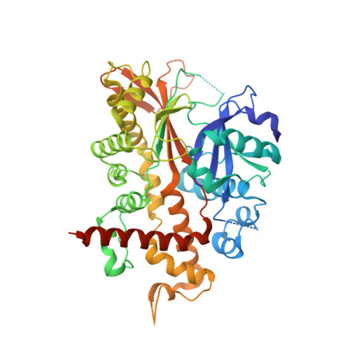
2XAM

Inositol 1,3,4,5,6-pentakisphosphate 2-kinase from A. thaliana in complex with ADP and IP6.


Experimental Data Snapshot

  • Method: X-RAY DIFFRACTION
  • Resolution: 2.20 Å
  • R-Value Free: 0.270 
  • R-Value Work: 0.227 
  • R-Value Observed: 0.229 

Starting Model: experimental
View more details

wwPDB Validation   3D Report Full Report


Ligand Structure Quality Assessment 


This is version 2.1 of the entry. See complete history


Literature

Inositol 1,3,4,5,6-Pentakisphosphate 2-Kinase is a Distant Ipk Member with a Singular Inositide Binding Site for Axial 2-Oh Recognition.

Gonzalez, B.Banos-Sanz, J.I.Villate, M.Brearley, C.A.Sanz-Aparicio, J.

(2010) Proc Natl Acad Sci U S A 107: 9608

  • DOI: https://doi.org/10.1073/pnas.0912979107
  • Primary Citation of Related Structures:  
    2XAL, 2XAM, 2XAN, 2XAO, 2XAR

  • PubMed Abstract: 

    Inositol phosphates (InsPs) are signaling molecules with multiple roles in cells. In particular (InsP(6)) is involved in mRNA export and editing or chromatin remodeling among other events. InsP(6) accumulates as mixed salts (phytate) in storage tissues of plants and plays a key role in their physiology. Human diets that are exclusively grain-based provide an excess of InsP(6) that, through chelation of metal ions, may have a detrimental effect on human health. Ins(1,3,4,5,6)P(5) 2-kinase (InsP(5) 2-kinase or Ipk1) catalyses the synthesis of InsP(6) from InsP(5) and ATP, and is the only enzyme that transfers a phosphate group to the axial 2-OH of the myo-inositide. We present the first structure for an InsP(5) 2-kinase in complex with both substrates and products. This enzyme presents a singular structural region for inositide binding that encompasses almost half of the protein. The key residues in substrate binding are identified, with Asp368 being responsible for recognition of the axial 2-OH. This study sheds light on the unique molecular mechanism for the synthesis of the precursor of inositol pyrophosphates.


  • Organizational Affiliation

    Grupo de Cristalografía Macromolecular y Biología Estructural, Instituto de Química-Física Rocasolano, Consejo Superior de Investigaciones Científicas, Serrano 119, 28006 Madrid, Spain. [email protected]


Macromolecules
Find similar proteins by:  (by identity cutoff)  |  3D Structure
Entity ID: 1
MoleculeChains Sequence LengthOrganismDetailsImage
INOSITOL-PENTAKISPHOSPHATE 2-KINASE
A, B
451Arabidopsis thalianaMutation(s): 0 
EC: 2.7.1.158
UniProt
Find proteins for Q93YN9 (Arabidopsis thaliana)
Explore Q93YN9 
Go to UniProtKB:  Q93YN9
Entity Groups  
Sequence Clusters30% Identity50% Identity70% Identity90% Identity95% Identity100% Identity
UniProt GroupQ93YN9
Sequence Annotations
Expand
  • Reference Sequence
Small Molecules
Ligands 4 Unique
IDChains Name / Formula / InChI Key2D Diagram3D Interactions
IHP
Query on IHP

Download Ideal Coordinates CCD File 
C [auth A],
H [auth B]
INOSITOL HEXAKISPHOSPHATE
C6 H18 O24 P6
IMQLKJBTEOYOSI-GPIVLXJGSA-N
ADP
Query on ADP

Download Ideal Coordinates CCD File 
D [auth A],
I [auth B]
ADENOSINE-5'-DIPHOSPHATE
C10 H15 N5 O10 P2
XTWYTFMLZFPYCI-KQYNXXCUSA-N
ZN
Query on ZN

Download Ideal Coordinates CCD File 
E [auth A],
J [auth B]
ZINC ION
Zn
PTFCDOFLOPIGGS-UHFFFAOYSA-N
MG
Query on MG

Download Ideal Coordinates CCD File 
F [auth A],
G [auth A],
K [auth B],
L [auth B]
MAGNESIUM ION
Mg
JLVVSXFLKOJNIY-UHFFFAOYSA-N
Experimental Data & Validation

Experimental Data

  • Method: X-RAY DIFFRACTION
  • Resolution: 2.20 Å
  • R-Value Free: 0.270 
  • R-Value Work: 0.227 
  • R-Value Observed: 0.229 
  • Space Group: P 21 21 21
Unit Cell:
Length ( Å )Angle ( ˚ )
a = 58.124α = 90
b = 113.591β = 90
c = 142.478γ = 90
Software Package:
Software NamePurpose
REFMACrefinement
MOSFLMdata reduction
CCP4data scaling
MOLREPphasing

Structure Validation

View Full Validation Report


Ligand Structure Quality Assessment 


Entry History 

Deposition Data

Revision History  (Full details and data files)

  • Version 1.0: 2010-05-19
    Type: Initial release
  • Version 1.1: 2011-07-13
    Changes: Advisory, Version format compliance
  • Version 1.2: 2012-02-29
    Changes: Other
  • Version 1.3: 2012-03-07
    Changes: Other
  • Version 1.4: 2016-12-21
    Changes: Structure summary
  • Version 1.5: 2018-03-07
    Changes: Source and taxonomy
  • Version 1.6: 2018-06-27
    Changes: Data collection, Derived calculations
  • Version 2.0: 2020-10-07
    Changes: Atomic model, Data collection, Derived calculations, Non-polymer description, Other, Structure summary
  • Version 2.1: 2023-12-20
    Changes: Data collection, Database references, Refinement description