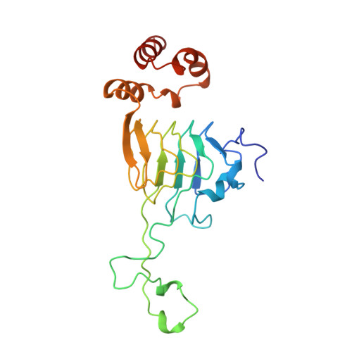
2XAT

COMPLEX OF THE HEXAPEPTIDE XENOBIOTIC ACETYLTRANSFERASE WITH CHLORAMPHENICOL AND DESULFO-COENZYME A


Experimental Data Snapshot

  • Method: X-RAY DIFFRACTION
  • Resolution: 3.20 Å
  • R-Value Free: 0.255 
  • R-Value Work: 0.213 
  • R-Value Observed: 0.213 

wwPDB Validation   3D Report Full Report


This is version 1.3 of the entry. See complete history


Literature

Structure of the hexapeptide xenobiotic acetyltransferase from Pseudomonas aeruginosa.

Beaman, T.W.Sugantino, M.Roderick, S.L.

(1998) Biochemistry 37: 6689-6696

  • DOI: https://doi.org/10.1021/bi980106v
  • Primary Citation of Related Structures:  
    1XAT, 2XAT

  • PubMed Abstract: 

    The crystal structure of the xenobiotic acetyltransferase from Pseudomonas aeruginosa PA103 (PaXAT) has been determined, as well as that of its complex with the substrate chloramphenicol and the cofactor analogue desulfo-coenzyme A. PaXAT is a member of the large hexapeptide acyltransferase family of enzymes that display tandem repeated copies of a six-residue hexapeptide repeat sequence motif encoding a left-handed parallel beta helix (L betaH) structural domain. The xenobiotic acetyltransferase class of hexapeptide acyltransferases is composed of microbial enzymes that utilize acetyl-CoA to acylate a variety of hydroxyl-bearing acceptors. The active site of trimeric PaXAT is a short tunnel into which chloramphenicol and the cofactor analogue desulfo-CoA project from opposite ends. This tunnel is formed by the flat parallel beta sheets of two separate L betaH domains and an extended 39-residue loop. His 79 of the extended loop forms hydrogen bonds from its imidazole NE2 atom to the 3-hydroxyl group of chloramphenicol and from its ND1 group to the peptide oxygen of Thr 86. The interactions of this histidine residue are similar to those found in the structurally unrelated type III chloramphenicol acetyltransferase and suggest that His 79 of PaXAT may be similarly positioned and tautomerically stabilized to serve as a general base catalyst.


  • Organizational Affiliation

    Department of Biochemistry, Albert Einstein College of Medicine, Bronx, New York 10461, USA.


Macromolecules
Find similar proteins by:  (by identity cutoff)  |  3D Structure
Entity ID: 1
MoleculeChains Sequence LengthOrganismDetailsImage
XENOBIOTIC ACETYLTRANSFERASE212Pseudomonas aeruginosaMutation(s): 0 
EC: 2.3.1.28
UniProt
Find proteins for P26841 (Pseudomonas aeruginosa (strain ATCC 15692 / DSM 22644 / CIP 104116 / JCM 14847 / LMG 12228 / 1C / PRS 101 / PAO1))
Explore P26841 
Go to UniProtKB:  P26841
Entity Groups  
Sequence Clusters30% Identity50% Identity70% Identity90% Identity95% Identity100% Identity
UniProt GroupP26841
Sequence Annotations
Expand
  • Reference Sequence
Small Molecules
Ligands 2 Unique
IDChains Name / Formula / InChI Key2D Diagram3D Interactions
DCA
Query on DCA

Download Ideal Coordinates CCD File 
C [auth A]DESULFO-COENZYME A
C21 H36 N7 O16 P3
ILWZMFJBPIYQKW-IBOSZNHHSA-N
CLM
Query on CLM

Download Ideal Coordinates CCD File 
B [auth A]CHLORAMPHENICOL
C11 H12 Cl2 N2 O5
WIIZWVCIJKGZOK-RKDXNWHRSA-N
Experimental Data & Validation

Experimental Data

  • Method: X-RAY DIFFRACTION
  • Resolution: 3.20 Å
  • R-Value Free: 0.255 
  • R-Value Work: 0.213 
  • R-Value Observed: 0.213 
  • Space Group: P 41 3 2
Unit Cell:
Length ( Å )Angle ( ˚ )
a = 154.8α = 90
b = 154.8β = 90
c = 154.8γ = 90
Software Package:
Software NamePurpose
PHASESphasing
X-PLORmodel building
X-PLORrefinement
XENGENdata reduction
XENGENdata scaling
X-PLORphasing

Structure Validation

View Full Validation Report


Entry History 

Deposition Data

Revision History  (Full details and data files)

  • Version 1.0: 1998-06-17
    Type: Initial release
  • Version 1.1: 2008-03-25
    Changes: Version format compliance
  • Version 1.2: 2011-07-13
    Changes: Derived calculations, Version format compliance
  • Version 1.3: 2024-02-21
    Changes: Data collection, Database references, Derived calculations