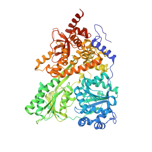
2XAU

Crystal structure of the Prp43p DEAH-box RNA helicase in complex with ADP


Experimental Data Snapshot

  • Method: X-RAY DIFFRACTION
  • Resolution: 1.90 Å
  • R-Value Free: 0.224 
  • R-Value Work: 0.184 
  • R-Value Observed: 0.186 

wwPDB Validation   3D Report Full Report


Ligand Structure Quality Assessment 


This is version 1.5 of the entry. See complete history


Literature

Prp43P Contains a Processive Helicase Structural Architecture with a Specific Regulatory Domain.

Walbott, H.Mouffok, S.Capeyrou, R.Lebaron, S.Humbert, O.Van Tilbeurgh, H.Henry, Y.Leulliot, N.

(2010) EMBO J 29: 2194

  • DOI: https://doi.org/10.1038/emboj.2010.102
  • Primary Citation of Related Structures:  
    2XAU

  • PubMed Abstract: 

    The DEAH/RNA helicase A (RHA) helicase family comprises proteins involved in splicing, ribosome biogenesis and transcription regulation. We report the structure of yeast Prp43p, a DEAH/RHA helicase remarkable in that it functions in both splicing and ribosome biogenesis. Prp43p displays a novel structural architecture with an unforeseen homology with the Ski2-like Hel308 DNA helicase. Together with the presence of a beta-hairpin in the second RecA-like domain, Prp43p contains all the structural elements of a processive helicase. Moreover, our structure reveals that the C-terminal domain contains an oligonucleotide/oligosaccharide-binding (OB)-fold placed at the entrance of the putative nucleic acid cavity. Deletion or mutations of this domain decrease the affinity of Prp43p for RNA and severely reduce Prp43p ATPase activity in the presence of RNA. We also show that this domain constitutes the binding site for the G-patch-containing domain of Pfa1p. We propose that the C-terminal domain, specific to DEAH/RHA helicases, is a central player in the regulation of helicase activity by binding both RNA and G-patch domain proteins.


  • Organizational Affiliation

    Institut de Biochimie et de Biophysique Moléculaire et Cellulaire, Université de Paris-Sud, CNRS-UMR8619, IFR115, Orsay Cedex, France.


Macromolecules
Find similar proteins by:  (by identity cutoff)  |  3D Structure
Entity ID: 1
MoleculeChains Sequence LengthOrganismDetailsImage
PRE-MRNA-SPLICING FACTOR ATP-DEPENDENT RNA HELICASE PRP43
A, B
773Saccharomyces cerevisiaeMutation(s): 0 
EC: 3.6.1 (PDB Primary Data), 3.6.4.13 (UniProt)
UniProt
Find proteins for P53131 (Saccharomyces cerevisiae (strain ATCC 204508 / S288c))
Explore P53131 
Go to UniProtKB:  P53131
Entity Groups  
Sequence Clusters30% Identity50% Identity70% Identity90% Identity95% Identity100% Identity
UniProt GroupP53131
Sequence Annotations
Expand
  • Reference Sequence
Small Molecules
Ligands 5 Unique
IDChains Name / Formula / InChI Key2D Diagram3D Interactions
ADP
Query on ADP

Download Ideal Coordinates CCD File 
C [auth A],
H [auth B]
ADENOSINE-5'-DIPHOSPHATE
C10 H15 N5 O10 P2
XTWYTFMLZFPYCI-KQYNXXCUSA-N
GOL
Query on GOL

Download Ideal Coordinates CCD File 
G [auth A],
L [auth B]
GLYCEROL
C3 H8 O3
PEDCQBHIVMGVHV-UHFFFAOYSA-N
ACT
Query on ACT

Download Ideal Coordinates CCD File 
F [auth A],
K [auth B]
ACETATE ION
C2 H3 O2
QTBSBXVTEAMEQO-UHFFFAOYSA-M
NI
Query on NI

Download Ideal Coordinates CCD File 
E [auth A],
J [auth B]
NICKEL (II) ION
Ni
VEQPNABPJHWNSG-UHFFFAOYSA-N
MG
Query on MG

Download Ideal Coordinates CCD File 
D [auth A],
I [auth B]
MAGNESIUM ION
Mg
JLVVSXFLKOJNIY-UHFFFAOYSA-N
Experimental Data & Validation

Experimental Data

  • Method: X-RAY DIFFRACTION
  • Resolution: 1.90 Å
  • R-Value Free: 0.224 
  • R-Value Work: 0.184 
  • R-Value Observed: 0.186 
  • Space Group: P 32 2 1
Unit Cell:
Length ( Å )Angle ( ˚ )
a = 117.551α = 90
b = 117.551β = 90
c = 254.605γ = 120
Software Package:
Software NamePurpose
PHENIXrefinement
MOSFLMdata reduction
SCALAdata scaling
autoSHARPphasing

Structure Validation

View Full Validation Report


Ligand Structure Quality Assessment 


Entry History 

Deposition Data

Revision History  (Full details and data files)

  • Version 1.0: 2010-06-09
    Type: Initial release
  • Version 1.1: 2011-05-08
    Changes: Version format compliance
  • Version 1.2: 2011-07-13
    Changes: Version format compliance
  • Version 1.3: 2019-01-30
    Changes: Data collection, Experimental preparation, Other
  • Version 1.4: 2019-02-06
    Changes: Data collection, Experimental preparation
  • Version 1.5: 2024-05-08
    Changes: Data collection, Database references, Derived calculations, Other