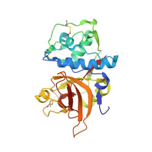
Halogen Bonding at the Active Sites of Human Cathepsin L and Mek1 Kinase: Efficient Interactions in Different Environments.
Hardegger, L.A., Kuhn, B., Spinnler, B., Anselm, L., Ecabert, R., Stihle, M., Gsell, B., Thoma, R., Diez, J., Benz, J.M., Plancher, J., Hartmann, G., Isshiki, Y., Morikami, K., Shimma, N., Haap, W., Banner, D.W., Diederich, F.(2011) ChemMedChem 6: 2048
- PubMed: 21898833
- DOI: https://doi.org/10.1002/cmdc.201100353
- Primary Citation of Related Structures:
2YJ2, 2YJ8, 2YJ9, 2YJB, 2YJC - PubMed Abstract:
In two series of small-molecule ligands, one inhibiting human cathepsin L (hcatL) and the other MEK1 kinase, biological affinities were found to strongly increase when an aryl ring of the inhibitors is substituted with the larger halogens Cl, Br, and I, but to decrease upon F substitution. X-ray co-crystal structure analyses revealed that the higher halides engage in halogen bonding (XB) with a backbone C=O in the S3 pocket of hcatL and in a back pocket of MEK1. While the S3 pocket is located at the surface of the enzyme, which provides a polar environment, the back pocket in MEK1 is deeply buried in the protein and is of pronounced apolar character. This study analyzes environmental effects on XB in protein-ligand complexes. It is hypothesized that energetic gains by XB are predominantly not due to water replacements but originate from direct interactions between the XB donor (Caryl-X) and the XB acceptor (C=O) in the correct geometry. New X-ray co-crystal structures in the same crystal form (space group P2(1)2(1)2(1)) were obtained for aryl chloride, bromide, and iodide ligands bound to hcatL. These high-resolution structures reveal that the backbone C=O group of Gly61 in most hcatL co-crystal structures maintains water solvation while engaging in XB. An aryl-CF3-substituted ligand of hcatL with an unexpectedly high affinity was found to adopt the same binding geometry as the aryl halides, with the CF3 group pointing to the C=O group of Gly61 in the S3 pocket. In this case, a repulsive F2C-F⋅⋅⋅O=C contact apparently is energetically overcompensated by other favorable protein-ligand contacts established by the CF3 group.
Organizational Affiliation:
Laboratorium für Organische Chemie, ETH Zürich, Wolfgang-Pauli-Strasse 10, HCI, 8093 Zürich, Switzerland.


