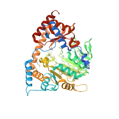
Crystal structure of LL-diaminopimelate aminotransferase from Arabidopsis thaliana: a recently discovered enzyme in the biosynthesis of L-lysine by plants and Chlamydia
Watanabe, N., Cherney, M.M., van Belkum, M.J., Marcus, S.L., Flegel, M.D., Clay, M.D., Deyholos, M.K., Vederas, J.C., James, M.N.(2007) J Mol Biol 371: 685-702
- PubMed: 17583737
- DOI: https://doi.org/10.1016/j.jmb.2007.05.061
- Primary Citation of Related Structures:
2Z1Z, 2Z20 - PubMed Abstract:
The essential biosynthetic pathway to l-Lysine in bacteria and plants is an attractive target for the development of new antibiotics or herbicides because it is absent in humans, who must acquire this amino acid in their diet. Plants use a shortcut of a bacterial pathway to l-Lysine in which the pyridoxal-5'-phosphate (PLP)-dependent enzyme ll-diaminopimelate aminotransferase (LL-DAP-AT) transforms l-tetrahydrodipicolinic acid (L-THDP) directly to LL-DAP. In addition, LL-DAP-AT was recently found in Chlamydia sp., suggesting that inhibitors of this enzyme may also be effective against such organisms. In order to understand the mechanism of this enzyme and to assist in the design of inhibitors, the three-dimensional crystal structure of LL-DAP-AT was determined at 1.95 A resolution. The cDNA sequence of LL-DAP-AT from Arabidopsis thaliana (AtDAP-AT) was optimized for expression in bacteria and cloned in Escherichia coli without its leader sequence but with a C-terminal hexahistidine affinity tag to aid protein purification. The structure of AtDAP-AT was determined using the multiple-wavelength anomalous dispersion (MAD) method with a seleno-methionine derivative. AtDAP-AT is active as a homodimer with each subunit having PLP in the active site. It belongs to the family of type I fold PLP-dependent enzymes. Comparison of the active site residues of AtDAP-AT and aspartate aminotransferases revealed that the PLP binding residues in AtDAP-AT are well conserved in both enzymes. However, Glu97* and Asn309* in the active site of AtDAP-AT are not found at similar positions in aspartate aminotransferases, suggesting that specific substrate recognition may require these residues from the other monomer. A malate-bound structure of AtDAP-AT allowed LL-DAP and L-glutamate to be modelled into the active site. These initial three-dimensional structures of LL-DAP-AT provide insight into its substrate specificity and catalytic mechanism.
Organizational Affiliation:
Group in Protein Structure and Function, Department of Biochemistry, University of Alberta, Edmonton, Alberta, Canada.


