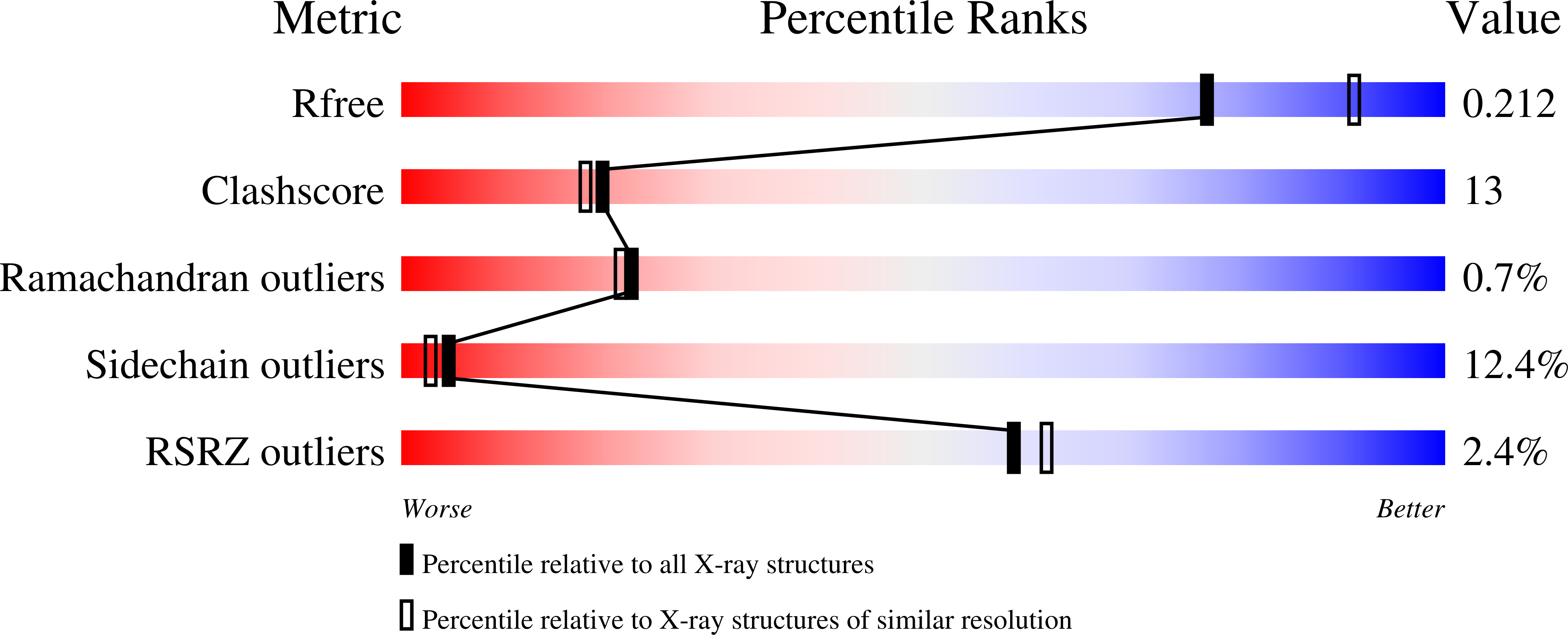
2ZFP

Thrombin Inibition


Experimental Data Snapshot

  • Method: X-RAY DIFFRACTION
  • Resolution: 2.25 Å
  • R-Value Free: 0.298 
  • R-Value Work: 0.176 
  • R-Value Observed: 0.180 

Starting Model: experimental
View more details

wwPDB Validation   3D Report Full Report


Ligand Structure Quality Assessment 


This is version 1.5 of the entry. See complete history


Literature

Think twice: understanding the high potency of bis(phenyl)methane inhibitors of thrombin

Baum, B.Muley, L.Heine, A.Smolinski, M.Hangauer, D.Klebe, G.

(2009) J Mol Biol 391: 552-564

  • DOI: https://doi.org/10.1016/j.jmb.2009.06.016
  • Primary Citation of Related Structures:  
    2ZC9, 2ZDA, 2ZFP, 2ZGX, 2ZO3, 3DHK, 3DUX, 3F68

  • PubMed Abstract: 

    Successful design of potent and selective protein inhibitors, in terms of structure-based drug design, strongly relies on the correct understanding of the molecular features determining the ligand binding to the target protein. We present a case study of serine protease inhibitors with a bis(phenyl)methane moiety binding into the S3 pocket. These inhibitors bind with remarkable potency to the active site of thrombin, the blood coagulation factor IIa. A combination of X-ray crystallography and isothermal titration calorimetry provides conclusive insights into the driving forces responsible for the surprisingly high potency of these inhibitors. Analysis of six well-resolved crystal structures (resolution 1.58-2.25 A) along with the thermodynamic data allows an explanation of the tight binding of the bis(phenyl)methane inhibitors. Interestingly, the two phenyl rings contribute to binding affinity for very different reasons - a fact that can only be elucidated by a structure-based approach. The first phenyl moiety occupies the hydrophobic S3 pocket, resulting in a mainly entropic advantage of binding. This observation is based on the displacement of structural water molecules from the S3 pocket that are observed in complexes with inhibitors that do not bind in the S3 pocket. The same classic hydrophobic effect cannot explain the enhanced binding affinity resulting from the attachment of the second, more solvent-exposed phenyl ring. For the bis(phenyl)methane inhibitors, an observed adaptive rotation of a glutamate residue adjacent to the S3 binding pocket attracted our attention. The rotation of this glutamate into salt-bridging distance with a lysine moiety correlates with an enhanced enthalpic contribution to binding for these highly potent thrombin binders. This explanation for the magnitude of the attractive force is confirmed by data retrieved by a Relibase search of several thrombin-inhibitor complexes deposited in the Protein Data Bank exhibiting similar molecular features. Special attention was attributed to putative changes in the protonation states of the interaction partners. For this purpose, two analogous inhibitors differing mainly in their potential to change the protonation state of a hydrogen-bond donor functionality were compared. Buffer dependencies of the binding enthalpy associated with complex formation could be traced by isothermal titration calorimetry, which revealed, along with analysis of the crystal structures (resolution 1.60 and 1.75 A), that a virtually compensating proton interchange between enzyme, inhibitor and buffer is responsible for the observed buffer-independent thermodynamic signatures.


  • Organizational Affiliation

    Department of Pharmaceutical Chemistry, Philipps-University Marburg, 35032 Marburg, Germany.


Macromolecules
Find similar proteins by:  (by identity cutoff)  |  3D Structure
Entity ID: 1
MoleculeChains Sequence LengthOrganismDetailsImage
Thrombin light chainA [auth L]36Homo sapiensMutation(s): 0 
EC: 3.4.21.5
UniProt & NIH Common Fund Data Resources
Find proteins for P00734 (Homo sapiens)
Explore P00734 
Go to UniProtKB:  P00734
PHAROS:  P00734
GTEx:  ENSG00000180210 
Entity Groups  
Sequence Clusters30% Identity50% Identity70% Identity90% Identity95% Identity100% Identity
UniProt GroupP00734
Sequence Annotations
Expand
  • Reference Sequence
Find similar proteins by:  (by identity cutoff)  |  3D Structure
Entity ID: 2
MoleculeChains Sequence LengthOrganismDetailsImage
Thrombin heavy chainB [auth H]259Homo sapiensMutation(s): 0 
EC: 3.4.21.5
UniProt & NIH Common Fund Data Resources
Find proteins for P00734 (Homo sapiens)
Explore P00734 
Go to UniProtKB:  P00734
PHAROS:  P00734
GTEx:  ENSG00000180210 
Entity Groups  
Sequence Clusters30% Identity50% Identity70% Identity90% Identity95% Identity100% Identity
UniProt GroupP00734
Sequence Annotations
Expand
  • Reference Sequence

Find similar proteins by:  Sequence   |   3D Structure  

Entity ID: 3
MoleculeChains Sequence LengthOrganismDetailsImage
Hirudin variant-1C [auth I]11N/AMutation(s): 0 
UniProt
Find proteins for P01050 (Hirudo medicinalis)
Explore P01050 
Go to UniProtKB:  P01050
Entity Groups  
Sequence Clusters30% Identity50% Identity70% Identity90% Identity95% Identity100% Identity
UniProt GroupP01050
Sequence Annotations
Expand
  • Reference Sequence
Small Molecules
Modified Residues  1 Unique
IDChains TypeFormula2D DiagramParent
TYS
Query on TYS
C [auth I]L-PEPTIDE LINKINGC9 H11 N O6 STYR
Binding Affinity Annotations 
IDSourceBinding Affinity
19U PDBBind:  2ZFP Ki: 6800 (nM) from 1 assay(s)
BindingDB:  2ZFP Ki: min: 6810, max: 1.30e+4 (nM) from 2 assay(s)
Biologically Interesting Molecules (External Reference) 1 Unique
Experimental Data & Validation

Experimental Data

  • Method: X-RAY DIFFRACTION
  • Resolution: 2.25 Å
  • R-Value Free: 0.298 
  • R-Value Work: 0.176 
  • R-Value Observed: 0.180 
  • Space Group: C 1 2 1
Unit Cell:
Length ( Å )Angle ( ˚ )
a = 70.5α = 90
b = 71.3β = 100.6
c = 72.9γ = 90
Software Package:
Software NamePurpose
SHELXmodel building
SHELXL-97refinement
CrystalCleardata collection
HKL-2000data reduction
HKL-2000data scaling
CNSphasing

Structure Validation

View Full Validation Report


Ligand Structure Quality Assessment 


Entry History 

Deposition Data

Revision History  (Full details and data files)

  • Version 1.0: 2008-12-16
    Type: Initial release
  • Version 1.1: 2011-07-13
    Changes: Atomic model, Database references, Derived calculations, Non-polymer description, Structure summary, Version format compliance
  • Version 1.2: 2012-01-18
    Changes: Non-polymer description
  • Version 1.3: 2012-12-12
    Changes: Other
  • Version 1.4: 2023-11-01
    Changes: Data collection, Database references, Derived calculations, Refinement description
  • Version 1.5: 2023-11-15
    Changes: Data collection