Understanding substrate misrecognition of hydrogen peroxide dependent cytochrome P450 from Bacillus subtilis.
Shoji, O., Fujishiro, T., Nagano, S., Tanaka, S., Hirose, T., Shiro, Y., Watanabe, Y.(2010) J Biol Inorg Chem
- PubMed: 20697922
- DOI: https://doi.org/10.1007/s00775-010-0692-4
- Primary Citation of Related Structures:
2ZQJ, 2ZQX - PubMed Abstract:
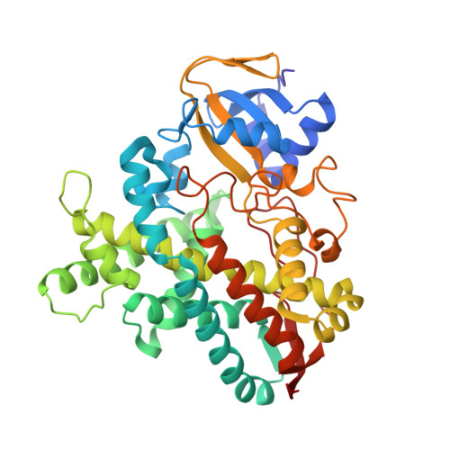
Cytochrome P450(BSβ), a H(2)O(2)-dependent cytochrome P450 catalyzing the hydroxylation of long-alkyl-chain fatty acids, lacks the general acid-base residue around the heme, which is indispensable for the efficient generation of the active species using H(2)O(2). On the basis of the crystal structure of the palmitic acid bound form of cytochrome P450(BSβ), it was suggested that the role of the general acid-base function was provided by the carboxylate group of fatty acids. The participation of the carboxylate group of the substrate was supported by the fact that cytochrome P450(BSβ) can catalyze oxidations of nonnatural substrates such as styrene and ethylbenzene in the presence of a series of short-alkyl-chain carboxylic acids as a dummy molecule of fatty acid. We refer to a series of short-alkyl-chain carboxylic acids as a "decoy molecule". As shown here, we have clarified the crystal structure of the decoy-molecule-bound form and elucidated that the location of its carboxylate group is virtually the same as that of palmitic acid in the heme cavity, indicating that the carboxylate group of the decoy molecule serves as the general acid-base catalyst. This result further confirms that the role of the acid-base function is satisfied by the carboxylate group of the substrates. In addition, the structure analysis of the substrate-free form has clarified that no remarkable structural change is induced by the binding of the decoy molecule as well as fatty acid. Consequently, whether the carboxylate group is positioned in the active site provides the switching mechanism of the catalytic cycle of cytochrome P450(BSβ).
Organizational Affiliation:
Department of Chemistry, Graduate School of Science, Nagoya University, Furo-cho, Chikusa-ku, Nagoya, 464-8602, Japan.


