2ZUV

Crystal structure of Galacto-N-biose/Lacto-N-biose I phosphorylase in complex with GlcNAc, Ethylene glycol, and nitrate


Experimental Data Snapshot

  • Method: X-RAY DIFFRACTION
  • Resolution: 1.85 Å
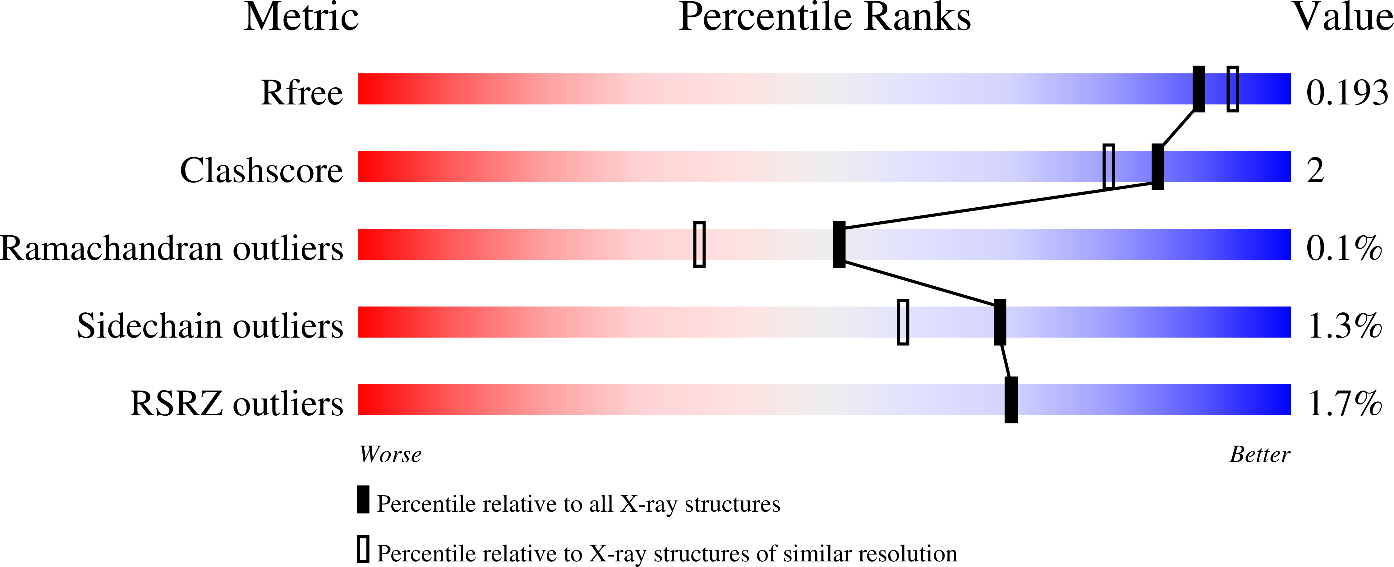
  • R-Value Free: 0.192 
  • R-Value Work: 0.149 
  • R-Value Observed: 0.151 

Starting Model: experimental
View more details

wwPDB Validation   3D Report Full Report


Ligand Structure Quality Assessment 


This is version 2.2 of the entry. See complete history


Literature

The crystal structure of galacto-N-biose/lacto-N-biose I phosphorylase: A large deformation of a tim barrel scaffold

Hidaka, M.Nishimoto, M.Kitaoka, M.Wakagi, T.Shoun, H.Fushinobu, S.

(2009) J Biol Chem 284: 7273-7283

  • DOI: https://doi.org/10.1074/jbc.M808525200
  • Primary Citation of Related Structures:  
    2ZUS, 2ZUT, 2ZUU, 2ZUV, 2ZUW

  • PubMed Abstract: 

    Galacto-N-biose/lacto-N-biose I phosphorylase (GLNBP) from Bifidobacterium longum, a key enzyme for intestinal growth, phosphorolyses galacto-N-biose and lacto-N-biose I with anomeric inversion. GLNBP homologues are often found in human pathogenic and commensal bacteria, and their substrate specificities potentially define the nutritional acquisition ability of these microbes in their habitat. We report the crystal structures of GLNBP in five different ligand-binding forms. This is the first three-dimensional structure of glycoside hydrolase (GH) family 112. The GlcNAc- and GalNAc-bound forms provide structural insights into distinct substrate preferences of GLNBP and its homologues from pathogens. The catalytic domain consists of a partially broken TIM barrel fold that is structurally similar to a thermophilic beta-galactosidase, strongly supporting the current classification of GLNBP homologues as one of the GH families. Anion binding induces a large conformational change by rotating a half-unit of the barrel. This is an unusual example of molecular adaptation of a TIM barrel scaffold to substrates.


  • Organizational Affiliation

    Department of Biotechnology, University of Tokyo, 1-1-1 Yayoi, Bunkyo-ku, Tokyo 113-8657, Japan.


Macromolecules
Find similar proteins by:  (by identity cutoff)  |  3D Structure
Entity ID: 1
MoleculeChains Sequence LengthOrganismDetailsImage
Lacto-N-biose phosphorylase
A, B
759Bifidobacterium longumMutation(s): 0 
Gene Names: lnpA
EC: 2.4.1.211
UniProt
Find proteins for E8MF13 (Bifidobacterium longum subsp. longum (strain ATCC 15707 / DSM 20219 / JCM 1217 / NCTC 11818 / E194b))
Explore E8MF13 
Go to UniProtKB:  E8MF13
Entity Groups  
Sequence Clusters30% Identity50% Identity70% Identity90% Identity95% Identity100% Identity
UniProt GroupE8MF13
Sequence Annotations
Expand
  • Reference Sequence
Small Molecules
Ligands 4 Unique
IDChains Name / Formula / InChI Key2D Diagram3D Interactions
NDG
Query on NDG

Download Ideal Coordinates CCD File 
C [auth A],
O [auth B]
2-acetamido-2-deoxy-alpha-D-glucopyranose
C8 H15 N O6
OVRNDRQMDRJTHS-PVFLNQBWSA-N
EDO
Query on EDO

Download Ideal Coordinates CCD File 
G [auth A]
H [auth A]
I [auth A]
J [auth A]
K [auth A]
G [auth A],
H [auth A],
I [auth A],
J [auth A],
K [auth A],
R [auth B],
S [auth B],
T [auth B],
U [auth B]
1,2-ETHANEDIOL
C2 H6 O2
LYCAIKOWRPUZTN-UHFFFAOYSA-N
NO3
Query on NO3

Download Ideal Coordinates CCD File 
D [auth A],
E [auth A],
F [auth A],
P [auth B],
Q [auth B]
NITRATE ION
N O3
NHNBFGGVMKEFGY-UHFFFAOYSA-N
MG
Query on MG

Download Ideal Coordinates CCD File 
L [auth A]
M [auth A]
N [auth A]
V [auth B]
W [auth B]
L [auth A],
M [auth A],
N [auth A],
V [auth B],
W [auth B],
X [auth B],
Y [auth B],
Z [auth B]
MAGNESIUM ION
Mg
JLVVSXFLKOJNIY-UHFFFAOYSA-N
Experimental Data & Validation

Experimental Data

  • Method: X-RAY DIFFRACTION
  • Resolution: 1.85 Å
  • R-Value Free: 0.192 
  • R-Value Work: 0.149 
  • R-Value Observed: 0.151 
  • Space Group: C 1 2 1
Unit Cell:
Length ( Å )Angle ( ˚ )
a = 204.043α = 90
b = 68.02β = 108.79
c = 116.047γ = 90
Software Package:
Software NamePurpose
DENZOdata reduction
SCALEPACKdata scaling
REFMACrefinement
PDB_EXTRACTdata extraction
HKL-2000data reduction
HKL-2000data scaling
MOLREPphasing

Structure Validation

View Full Validation Report


Ligand Structure Quality Assessment 


Entry History 

Deposition Data

Revision History  (Full details and data files)

  • Version 1.0: 2008-12-30
    Type: Initial release
  • Version 1.1: 2011-07-13
    Changes: Version format compliance
  • Version 1.2: 2017-10-11
    Changes: Advisory, Refinement description
  • Version 2.0: 2020-07-29
    Type: Remediation
    Reason: Carbohydrate remediation
    Changes: Atomic model, Data collection, Database references, Derived calculations, Structure summary
  • Version 2.1: 2024-03-13
    Changes: Advisory, Data collection, Database references, Structure summary
  • Version 2.2: 2024-04-03
    Changes: Refinement description