2ZVP

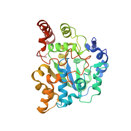
Crystal structure of mouse cytosolic sulfotransferase mSULT1D1 complex with PAP and p-nitrophenol


Experimental Data Snapshot

  • Method: X-RAY DIFFRACTION
  • Resolution: 1.30 Å
  • R-Value Free: 0.190 
  • R-Value Work: 0.175 
  • R-Value Observed: 0.175 

Starting Model: experimental
View more details

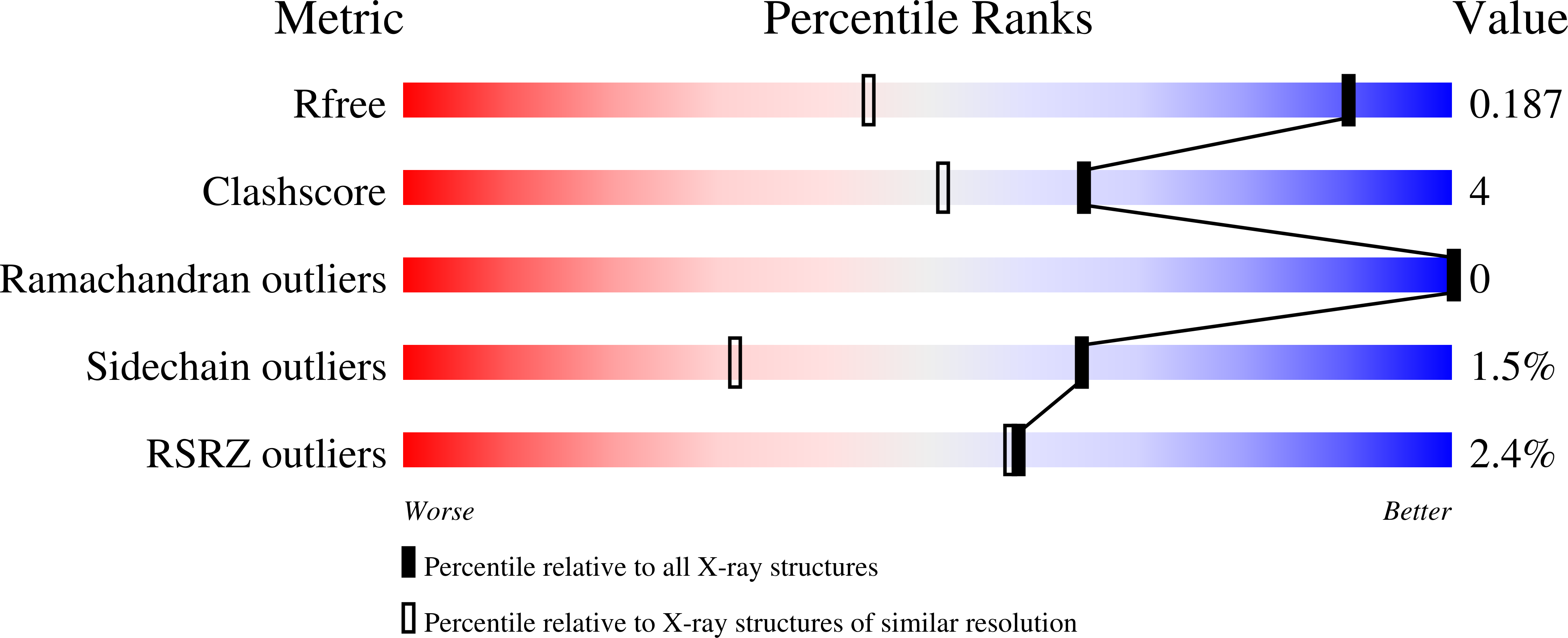
wwPDB Validation   3D Report Full Report


Ligand Structure Quality Assessment 


This is version 1.2 of the entry. See complete history


Literature

Structural basis for the broad range substrate specificity of a novel mouse cytosolic sulfotransferase--mSULT1D1

Teramoto, T.Sakakibara, Y.Liu, M.-C.Suiko, M.Kimura, M.Kakuta, Y.

(2009) Biochem Biophys Res Commun 379: 76-80

  • DOI: https://doi.org/10.1016/j.bbrc.2008.12.013
  • Primary Citation of Related Structures:  
    2ZVP, 2ZVQ

  • PubMed Abstract: 

    The mouse cytosolic sulfotransferase, mSULT1D1, catalyzes the sulfonation of a wide range of phenolic molecules including p-nitrophenol (pNP), alpha-naphthol (alphaNT), serotonin, as well as dopamine and its metabolites. To gain insight into the structural basis for its broad range substrate specificity, we solved two distinct ternary crystal structures of mSULT1D1, complexed with 3'-phosphoadenosine-5'-phosphate (PAP) plus pNP or PAP plus alphaNT. The structures revealed that the mSULT1D1 contains an L-shaped accepter-binding site which comprises 20 amino acid residues and four conserved water molecules. The shape of the accepter-binding site can be adjusted by conformational changes of two residues, Ile148 and Glu247, upon binding with respective substrates.


  • Organizational Affiliation

    Department of Systems Life Sciences, Laboratory of Structural Biology, Graduate School, Faculty of Agriculture, Kyushu University, Fukuoka, Japan.


Macromolecules
Find similar proteins by:  (by identity cutoff)  |  3D Structure
Entity ID: 1
MoleculeChains Sequence LengthOrganismDetailsImage
Tyrosine-ester sulfotransferaseA [auth X]295Mus musculusMutation(s): 0 
Gene Names: Sult1d1
EC: 2.8.2.9 (PDB Primary Data), 2.8.2 (UniProt)
UniProt
Find proteins for Q3UZZ6 (Mus musculus)
Explore Q3UZZ6 
Go to UniProtKB:  Q3UZZ6
Entity Groups  
Sequence Clusters30% Identity50% Identity70% Identity90% Identity95% Identity100% Identity
UniProt GroupQ3UZZ6
Sequence Annotations
Expand
  • Reference Sequence
Experimental Data & Validation

Experimental Data

  • Method: X-RAY DIFFRACTION
  • Resolution: 1.30 Å
  • R-Value Free: 0.190 
  • R-Value Work: 0.175 
  • R-Value Observed: 0.175 
  • Space Group: C 1 2 1
Unit Cell:
Length ( Å )Angle ( ˚ )
a = 156.509α = 90
b = 67.615β = 105.47
c = 42.721γ = 90
Software Package:
Software NamePurpose
REFMACrefinement
BBSdata collection
HKL-2000data reduction
HKL-2000data scaling
MOLREPphasing

Structure Validation

View Full Validation Report


Ligand Structure Quality Assessment 


Entry History 

Deposition Data

Revision History  (Full details and data files)

  • Version 1.0: 2008-12-30
    Type: Initial release
  • Version 1.1: 2011-07-13
    Changes: Non-polymer description, Version format compliance
  • Version 1.2: 2023-11-01
    Changes: Data collection, Database references, Refinement description