2DFK

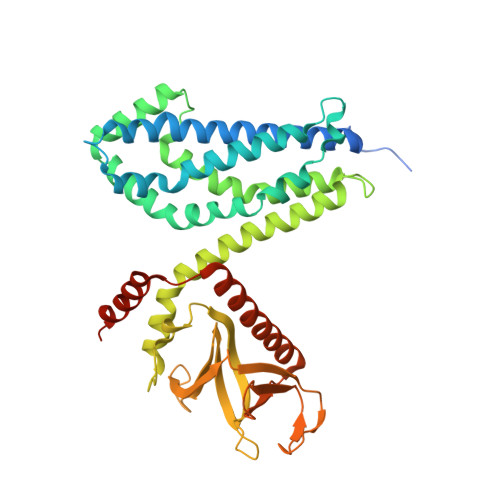
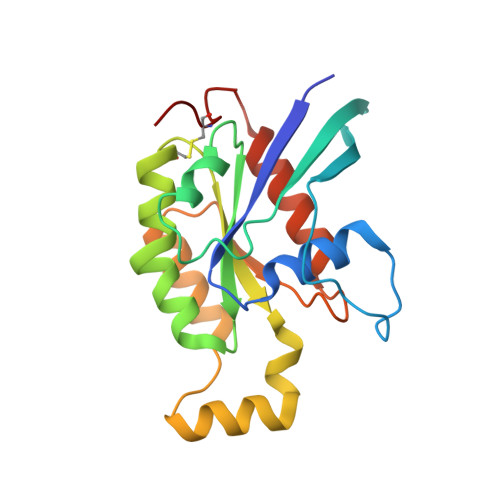
Crystal structure of the CDC42-Collybistin II complex


Experimental Data Snapshot

  • Method: X-RAY DIFFRACTION
  • Resolution: 2.15 Å
  • R-Value Free: 0.229 
  • R-Value Work: 0.180 
  • R-Value Observed: 0.183 

Starting Model: experimental
View more details

wwPDB Validation   3D Report Full Report


This is version 1.4 of the entry. See complete history


Literature

The Crystal Structure of Cdc42 in Complex with Collybistin II, a Gephyrin-interacting Guanine Nucleotide Exchange Factor.

Xiang, S.Kim, E.Y.Connelly, J.J.Nassar, N.Kirsch, J.Winking, J.Schwarz, G.Schindelin, H.

(2006) J Mol Biol 359: 35-46

  • DOI: https://doi.org/10.1016/j.jmb.2006.03.019
  • Primary Citation of Related Structures:  
    2DFK

  • PubMed Abstract: 

    The synaptic localization of ion channel receptors is essential for efficient synaptic transmission and the precise regulation of diverse neuronal functions. In the central nervous system, ion channel receptors reside in the postsynaptic membrane where they are juxtaposed to presynaptic terminals. For proper function, these ion channels have to be anchored to the cytoskeleton, and in the case of the inhibitory glycine and gamma-amino-butyric acid type A (GABA(A)) receptors this interaction is mediated by a gephyrin centered scaffold. Highlighting its central role in this receptor anchoring scaffold, gephyrin interacts with a number of proteins, including the neurospecific guanine nucleotide exchange factor collybistin. Collybistin belongs to the Dbl family of guanine nucleotide exchange factors, occurs in multiple splice variants, and is specific for Cdc42, a small GTPase belonging to the Rho family. The 2.3 Angstroms resolution crystal structure of the Cdc42-collybistin II complex reveals a novel conformation of the switch I region of Cdc42. It also provides the first direct observation of structural changes in the relative orientation of the Dbl-homology domain and the pleckstrin-homology domain in the same Dbl family protein. Biochemical data indicate that gephyrin negatively regulates collybistin activity.


  • Organizational Affiliation

    Department of Biochemistry, SUNY Stony Brook, NY 11794-5215, USA.


Macromolecules
Find similar proteins by:  (by identity cutoff)  |  3D Structure
Entity ID: 1
MoleculeChains Sequence LengthOrganismDetailsImage
collybistin II
A, C
402Rattus norvegicusMutation(s): 0 
UniProt
Find proteins for Q9QX73 (Rattus norvegicus)
Explore Q9QX73 
Go to UniProtKB:  Q9QX73
Entity Groups  
Sequence Clusters30% Identity50% Identity70% Identity90% Identity95% Identity100% Identity
UniProt GroupQ9QX73
Sequence Annotations
Expand
  • Reference Sequence
Find similar proteins by:  (by identity cutoff)  |  3D Structure
Entity ID: 2
MoleculeChains Sequence LengthOrganismDetailsImage
cell division cycle 42 isoform 1
B, D
194Homo sapiensMutation(s): 0 
EC: 3.6.5.2
Membrane Entity: Yes 
UniProt & NIH Common Fund Data Resources
Find proteins for P60953 (Homo sapiens)
Explore P60953 
Go to UniProtKB:  P60953
PHAROS:  P60953
GTEx:  ENSG00000070831 
Entity Groups  
Sequence Clusters30% Identity50% Identity70% Identity90% Identity95% Identity100% Identity
UniProt GroupP60953
Sequence Annotations
Expand
  • Reference Sequence
Small Molecules
Ligands 2 Unique
IDChains Name / Formula / InChI Key2D Diagram3D Interactions
SO4
Query on SO4

Download Ideal Coordinates CCD File 
H [auth B],
J [auth D]
SULFATE ION
O4 S
QAOWNCQODCNURD-UHFFFAOYSA-L
GOL
Query on GOL

Download Ideal Coordinates CCD File 
E [auth A]
F [auth A]
G [auth A]
I [auth B]
K [auth D]
E [auth A],
F [auth A],
G [auth A],
I [auth B],
K [auth D],
L [auth D]
GLYCEROL
C3 H8 O3
PEDCQBHIVMGVHV-UHFFFAOYSA-N
Experimental Data & Validation

Experimental Data

  • Method: X-RAY DIFFRACTION
  • Resolution: 2.15 Å
  • R-Value Free: 0.229 
  • R-Value Work: 0.180 
  • R-Value Observed: 0.183 
  • Space Group: P 21 21 21
Unit Cell:
Length ( Å )Angle ( ˚ )
a = 57.957α = 90
b = 147.499β = 90
c = 167.22γ = 90
Software Package:
Software NamePurpose
DENZOdata reduction
SCALEPACKdata scaling
MOLREPphasing
REFMACrefinement

Structure Validation

View Full Validation Report


Entry History 

Deposition Data

Revision History  (Full details and data files)

  • Version 1.0: 2006-05-02
    Type: Initial release
  • Version 1.1: 2008-04-30
    Changes: Version format compliance
  • Version 1.2: 2011-07-13
    Changes: Non-polymer description, Version format compliance
  • Version 1.3: 2023-10-25
    Changes: Data collection, Database references, Derived calculations, Refinement description
  • Version 1.4: 2024-10-30
    Changes: Structure summary