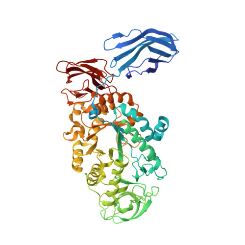
3BCD

Alpha-amylase B in complex with maltotetraose and alpha-cyclodextrin


Experimental Data Snapshot

  • Method: X-RAY DIFFRACTION
  • Resolution: 2.20 Å
  • R-Value Free: 0.240 
  • R-Value Work: 0.196 
  • R-Value Observed: 0.197 

Starting Model: experimental
View more details

wwPDB Validation   3D Report Full Report


This is version 2.1 of the entry. See complete history


Literature

Crystal Structure of the Polyextremophilic alpha-Amylase AmyB from Halothermothrix orenii: Details of a Productive Enzyme-Substrate Complex and an N Domain with a Role in Binding Raw Starch

Tan, T.-C.Mijts, B.N.Swaminathan, K.Patel, B.K.C.Divne, C.

(2008) J Mol Biol 378: 850-868

  • DOI: https://doi.org/10.1016/j.jmb.2008.02.041
  • Primary Citation of Related Structures:  
    3BC9, 3BCD, 3BCF

  • PubMed Abstract: 

    The gene for a membrane-bound, halophilic, and thermostable alpha-amylase, AmyB, from Halothermothrix orenii was cloned and sequenced. The crystal structure shows that, in addition to the typical domain organization of family 13 glycoside hydrolases, AmyB carries an additional N-terminal domain (N domain) that forms a large groove--the N-C groove--some 30 A away from the active site. The structure of AmyB with the inhibitor acarbose at 1.35 A resolution shows that a nonasaccharide has been synthesized through successive transglycosylation reactions of acarbose. Unexpectedly, in a complex of wild-type AmyB with alpha-cyclodextrin and maltoheptaose at 2.2 A resolution, a maltotetraose molecule is bound in subsites -1 to +3, spanning the cleavage point at -1/+1, with the -1 glucosyl residue present as a (2)S(o) skew boat. This wild-type AmyB complex was obtained in the presence of a large excess of substrate, a condition under which it is possible to capture Michaelis complexes, which may explain the observed binding across -1/+1 and ring distortion. We observe three methionine side chains that serve as "binding platforms" for glucosyl rings in AmyB, a seemingly rare occurrence in carbohydrate-binding proteins. The structures and results from the biochemical characterization of AmyB and AmyB lacking the N domain show that the N domain increases binding of the enzyme to raw starch. Furthermore, theoretical modeling suggests that the N-C groove can accommodate, spatially and chemically, large substrates such as A-starch.


  • Organizational Affiliation

    KTH School of Biotechnology, AlbaNova University Center, Roslagstullsbacken 21, SE-106 91 Stockholm, Sweden.


Macromolecules
Find similar proteins by:  (by identity cutoff)  |  3D Structure
Entity ID: 1
MoleculeChains Sequence LengthOrganismDetailsImage
Alpha amylase, catalytic region599Halothermothrix orenii H 168Mutation(s): 0 
Gene Names: amyb
EC: 3.2.1.1 (PDB Primary Data), 3.2.1.98 (UniProt)
UniProt
Find proteins for B8CZ54 (Halothermothrix orenii (strain H 168 / OCM 544 / DSM 9562))
Explore B8CZ54 
Go to UniProtKB:  B8CZ54
Entity Groups  
Sequence Clusters30% Identity50% Identity70% Identity90% Identity95% Identity100% Identity
UniProt GroupB8CZ54
Sequence Annotations
Expand
  • Reference Sequence
Oligosaccharides

Help

Entity ID: 2
MoleculeChains Length2D Diagram Glycosylation3D Interactions
alpha-D-glucopyranose-(1-4)-alpha-D-glucopyranose-(1-4)-alpha-D-glucopyranose-(1-4)-beta-D-glucopyranose
B
4N/A
Glycosylation Resources
GlyTouCan:  G69211UR
GlyCosmos:  G69211UR
GlyGen:  G69211UR
Entity ID: 3
MoleculeChains Length2D Diagram Glycosylation3D Interactions
alpha-D-glucopyranose-(1-4)-alpha-D-glucopyranose-(1-4)-alpha-D-glucopyranose-(1-4)-alpha-D-glucopyranose
C
4N/A
Glycosylation Resources
GlyTouCan:  G87171PZ
GlyCosmos:  G87171PZ
GlyGen:  G87171PZ
Entity ID: 4
MoleculeChains Length2D Diagram Glycosylation3D Interactions
Cyclohexakis-(1-4)-(alpha-D-glucopyranose)
D
6N/A
Glycosylation Resources
GlyTouCan:  G22307HL
GlyCosmos:  G22307HL
Small Molecules
Ligands 2 Unique
IDChains Name / Formula / InChI Key2D Diagram3D Interactions
CA
Query on CA

Download Ideal Coordinates CCD File 
E [auth A]
F [auth A]
G [auth A]
I [auth A]
J [auth A]
E [auth A],
F [auth A],
G [auth A],
I [auth A],
J [auth A],
K [auth A],
L [auth A]
CALCIUM ION
Ca
BHPQYMZQTOCNFJ-UHFFFAOYSA-N
NA
Query on NA

Download Ideal Coordinates CCD File 
H [auth A]SODIUM ION
Na
FKNQFGJONOIPTF-UHFFFAOYSA-N
Biologically Interesting Molecules (External Reference) 2 Unique
Experimental Data & Validation

Experimental Data

  • Method: X-RAY DIFFRACTION
  • Resolution: 2.20 Å
  • R-Value Free: 0.240 
  • R-Value Work: 0.196 
  • R-Value Observed: 0.197 
  • Space Group: C 1 2 1
Unit Cell:
Length ( Å )Angle ( ˚ )
a = 228.7α = 90
b = 78.05β = 98.9
c = 50.64γ = 90
Software Package:
Software NamePurpose
REFMACrefinement
MAR345data collection
XDSdata reduction
XSCALEdata scaling
REFMACphasing

Structure Validation

View Full Validation Report


Entry History 

Deposition Data

Revision History  (Full details and data files)

  • Version 1.0: 2008-04-22
    Type: Initial release
  • Version 1.1: 2011-07-13
    Changes: Advisory, Source and taxonomy, Version format compliance
  • Version 1.2: 2017-10-25
    Changes: Data collection, Refinement description
  • Version 2.0: 2020-07-29
    Type: Remediation
    Reason: Carbohydrate remediation
    Changes: Atomic model, Data collection, Derived calculations, Non-polymer description, Structure summary
  • Version 2.1: 2023-11-01
    Changes: Data collection, Database references, Refinement description, Structure summary