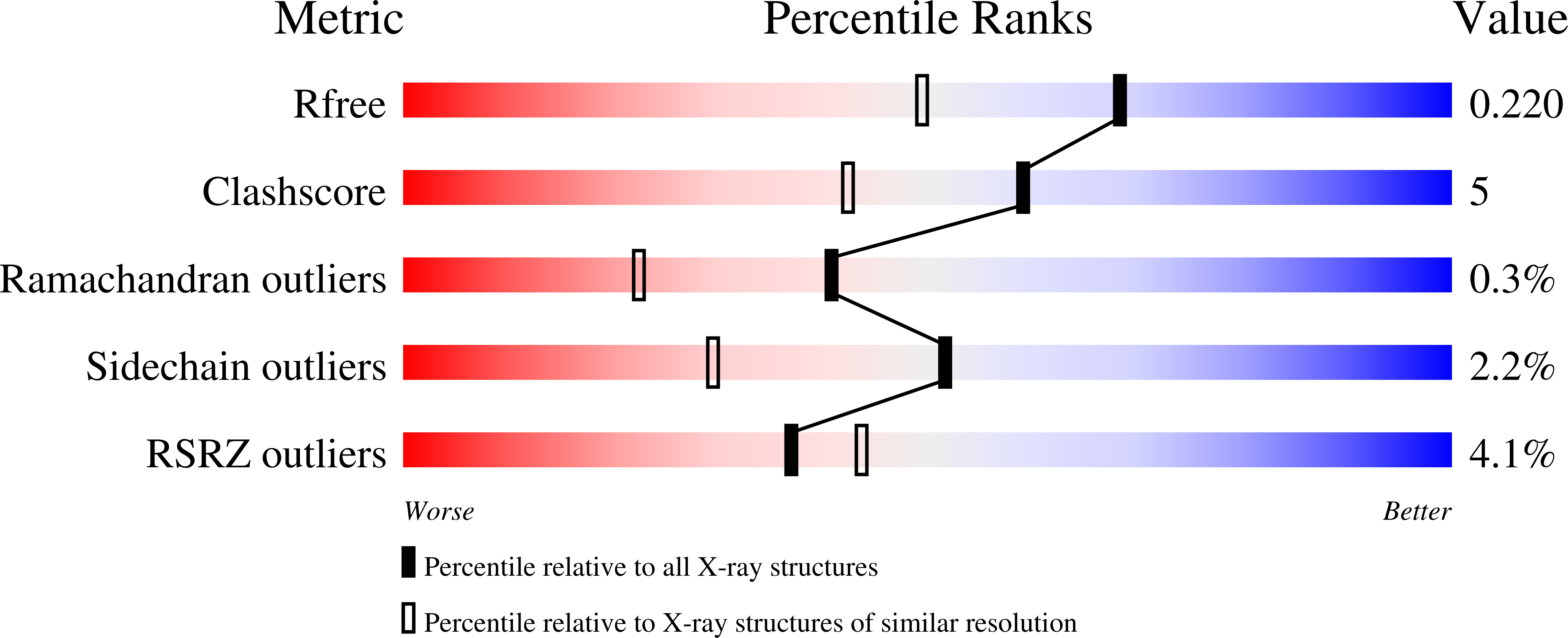
The crystal structure of [Fe]-hydrogenase reveals the geometry of the active site.
Shima, S., Pilak, O., Vogt, S., Schick, M., Stagni, M.S., Meyer-Klaucke, W., Warkentin, E., Thauer, R.K., Ermler, U.(2008) Science 321: 572-575
- PubMed: 18653896
- DOI: https://doi.org/10.1126/science.1158978
- Primary Citation of Related Structures:
3DAF, 3DAG - PubMed Abstract:
Biological formation and consumption of molecular hydrogen (H2) are catalyzed by hydrogenases, of which three phylogenetically unrelated types are known: [NiFe]-hydrogenases, [FeFe]-hydrogenases, and [Fe]-hydrogenase. We present a crystal structure of [Fe]-hydrogenase at 1.75 angstrom resolution, showing a mononuclear iron coordinated by the sulfur of cysteine 176, two carbon monoxide (CO) molecules, and the sp2-hybridized nitrogen of a 2-pyridinol compound with back-bonding properties similar to those of cyanide. The three-dimensional arrangement of the ligands is similar to that of thiolate, CO, and cyanide ligated to the low-spin iron in binuclear [NiFe]- and [FeFe]-hydrogenases, although the enzymes have evolved independently and the CO and cyanide ligands are not found in any other metalloenzyme. The related iron ligation pattern of hydrogenases exemplifies convergent evolution and presumably plays an essential role in H2 activation. This finding may stimulate the ongoing synthesis of catalysts that could substitute for platinum in applications such as fuel cells.
Organizational Affiliation:
Max-Planck-Institut für Terrestrische Mikrobiologie and Laboratorium für Mikrobiologie, Fachbereich Biologie, Philipps-Universität Marburg, Karl-von-Frisch-Strasse, D-35043 Marburg, Germany. [email protected]


