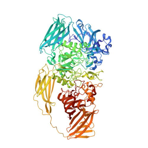
3DYO

E. coli (lacZ) beta-galactosidase (H418N) in complex with IPTG


Experimental Data Snapshot

  • Method: X-RAY DIFFRACTION
  • Resolution: 1.80 Å
  • R-Value Free: 0.241 
  • R-Value Work: 0.173 

Starting Model: experimental
View more details

wwPDB Validation   3D Report Full Report


Ligand Structure Quality Assessment 


This is version 1.5 of the entry. See complete history


Literature

Direct and indirect roles of His-418 in metal binding and in the activity of beta-galactosidase (E. coli).

Juers, D.H.Rob, B.Dugdale, M.L.Rahimzadeh, N.Giang, C.Lee, M.Matthews, B.W.Huber, R.E.

(2009) Protein Sci 18: 1281-1292

  • DOI: https://doi.org/10.1002/pro.140
  • Primary Citation of Related Structures:  
    3DYM, 3DYO, 3DYP, 3E1F

  • PubMed Abstract: 

    The active site of ss-galactosidase (E. coli) contains a Mg(2+) ion ligated by Glu-416, His-418 and Glu-461 plus three water molecules. A Na(+) ion binds nearby. To better understand the role of the active site Mg(2+) and its ligands, His-418 was substituted with Asn, Glu and Phe. The Asn-418 and Glu-418 variants could be crystallized and the structures were shown to be very similar to native enzyme. The Glu-418 variant showed increased mobility of some residues in the active site, which explains why the substitutions at the Mg(2+) site also reduce Na(+) binding affinity. The Phe variant had reduced stability, bound Mg(2+) weakly and could not be crystallized. All three variants have low catalytic activity due to large decreases in the degalactosylation rate. Large decreases in substrate binding affinity were also observed but transition state analogs bound as well or better than to native. The results indicate that His-418, together with the Mg(2+), modulate the central role of Glu-461 in binding and as a general acid/base catalyst in the overall catalytic mechanism. Glucose binding as an acceptor was also dramatically decreased, indicating that His-418 is very important for the formation of allolactose (the natural inducer of the lac operon).


  • Organizational Affiliation

    Instititute of Molecular Biology, Howard Hughes Medical Institute and Department of Physics, University of Oregon, Eugene, OR 97403-1229, USA.


Macromolecules
Find similar proteins by:  (by identity cutoff)  |  3D Structure
Entity ID: 1
MoleculeChains Sequence LengthOrganismDetailsImage
Beta-galactosidase
A, B, C, D
1,023Escherichia coli K-12Mutation(s): 1 
Gene Names: lacZb0344JW0335
EC: 3.2.1.23
UniProt
Find proteins for P00722 (Escherichia coli (strain K12))
Explore P00722 
Go to UniProtKB:  P00722
Entity Groups  
Sequence Clusters30% Identity50% Identity70% Identity90% Identity95% Identity100% Identity
UniProt GroupP00722
Sequence Annotations
Expand
  • Reference Sequence
Small Molecules
Ligands 4 Unique
IDChains Name / Formula / InChI Key2D Diagram3D Interactions
IPT
Query on IPT

Download Ideal Coordinates CCD File 
HC [auth C]
HD [auth C]
IC [auth C]
M [auth A]
MA [auth A]
HC [auth C],
HD [auth C],
IC [auth C],
M [auth A],
MA [auth A],
N [auth A],
PD [auth D],
QD [auth D],
RD [auth D],
WA [auth B],
XA [auth B],
YB [auth B]
1-methylethyl 1-thio-beta-D-galactopyranoside
C9 H18 O5 S
BPHPUYQFMNQIOC-NXRLNHOXSA-N
DMS
Query on DMS

Download Ideal Coordinates CCD File 
AA [auth A]
AB [auth B]
AD [auth C]
AE [auth D]
BA [auth A]
AA [auth A],
AB [auth B],
AD [auth C],
AE [auth D],
BA [auth A],
BB [auth B],
BD [auth C],
BE [auth D],
CA [auth A],
CB [auth B],
CD [auth C],
CE [auth D],
DA [auth A],
DB [auth B],
DD [auth C],
DE [auth D],
EA [auth A],
EB [auth B],
ED [auth C],
EE [auth D],
FA [auth A],
FB [auth B],
FD [auth C],
FE [auth D],
GA [auth A],
GB [auth B],
GD [auth C],
GE [auth D],
HA [auth A],
HB [auth B],
HE [auth D],
IA [auth A],
IB [auth B],
IE [auth D],
JA [auth A],
JB [auth B],
JC [auth C],
JE [auth D],
KA [auth A],
KB [auth B],
KC [auth C],
KE [auth D],
LA [auth A],
LB [auth B],
LC [auth C],
LE [auth D],
MB [auth B],
MC [auth C],
ME [auth D],
NB [auth B],
NC [auth C],
NE [auth D],
O [auth A],
OB [auth B],
OC [auth C],
OE [auth D],
P [auth A],
PB [auth B],
PC [auth C],
Q [auth A],
QB [auth B],
QC [auth C],
R [auth A],
RB [auth B],
RC [auth C],
S [auth A],
SB [auth B],
SC [auth C],
SD [auth D],
T [auth A],
TB [auth B],
TC [auth C],
TD [auth D],
U [auth A],
UB [auth B],
UC [auth C],
UD [auth D],
V [auth A],
VB [auth B],
VC [auth C],
VD [auth D],
W [auth A],
WB [auth B],
WC [auth C],
WD [auth D],
X [auth A],
XB [auth B],
XC [auth C],
XD [auth D],
Y [auth A],
YA [auth B],
YC [auth C],
YD [auth D],
Z [auth A],
ZA [auth B],
ZC [auth C],
ZD [auth D]
DIMETHYL SULFOXIDE
C2 H6 O S
IAZDPXIOMUYVGZ-UHFFFAOYSA-N
MG
Query on MG

Download Ideal Coordinates CCD File 
AC [auth C]
BC [auth C]
E [auth A]
F [auth A]
G [auth A]
AC [auth C],
BC [auth C],
E [auth A],
F [auth A],
G [auth A],
H [auth A],
ID [auth D],
JD [auth D],
KD [auth D],
NA [auth B],
OA [auth B],
PA [auth B],
QA [auth B],
ZB [auth C]
MAGNESIUM ION
Mg
JLVVSXFLKOJNIY-UHFFFAOYSA-N
NA
Query on NA

Download Ideal Coordinates CCD File 
CC [auth C]
DC [auth C]
EC [auth C]
FC [auth C]
GC [auth C]
CC [auth C],
DC [auth C],
EC [auth C],
FC [auth C],
GC [auth C],
I [auth A],
J [auth A],
K [auth A],
L [auth A],
LD [auth D],
MD [auth D],
ND [auth D],
OD [auth D],
RA [auth B],
SA [auth B],
TA [auth B],
UA [auth B],
VA [auth B]
SODIUM ION
Na
FKNQFGJONOIPTF-UHFFFAOYSA-N
Binding Affinity Annotations 
IDSourceBinding Affinity
IPT BindingDB:  3DYO Ki: 7.60e+4 (nM) from 1 assay(s)
PDBBind:  3DYO Ki: 5.00e+6 (nM) from 1 assay(s)
Experimental Data & Validation

Experimental Data

  • Method: X-RAY DIFFRACTION
  • Resolution: 1.80 Å
  • R-Value Free: 0.241 
  • R-Value Work: 0.173 
  • Space Group: P 21 21 21
Unit Cell:
Length ( Å )Angle ( ˚ )
a = 152.01α = 90
b = 162.52β = 90
c = 204.01γ = 90
Software Package:
Software NamePurpose
TNTrefinement
MOSFLMdata reduction
SCALAdata scaling
TNTphasing

Structure Validation

View Full Validation Report


Ligand Structure Quality Assessment 


Entry History 

Deposition Data

Revision History  (Full details and data files)

  • Version 1.0: 2008-10-28
    Type: Initial release
  • Version 1.1: 2011-07-13
    Changes: Version format compliance
  • Version 1.2: 2020-07-29
    Type: Remediation
    Reason: Carbohydrate remediation
    Changes: Data collection, Database references, Derived calculations, Structure summary
  • Version 1.3: 2021-07-28
    Changes: Refinement description, Structure summary
  • Version 1.4: 2021-10-20
    Changes: Database references
  • Version 1.5: 2023-08-30
    Changes: Data collection, Refinement description