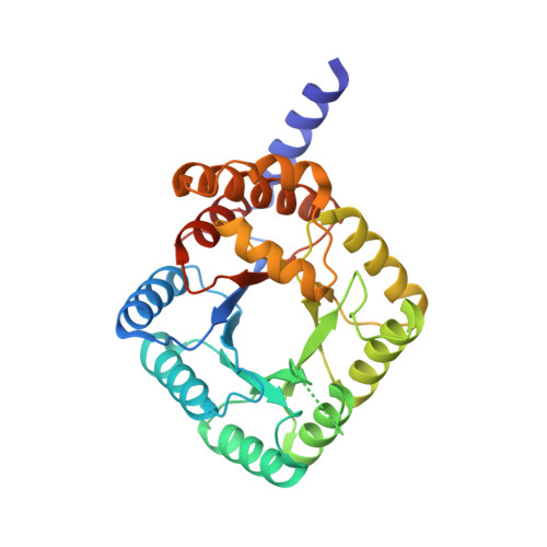
3FSU

Crystal Structure of Escherichia coli Methylenetetrahydrofolate Reductase Double Mutant Phe223LeuGlu28Gln complexed with methyltetrahydrofolate


Experimental Data Snapshot

  • Method: X-RAY DIFFRACTION
  • Resolution: 1.70 Å
  • R-Value Free: 0.220 
  • R-Value Work: 0.197 
  • R-Value Observed: 0.198 

Starting Model: experimental
View more details

wwPDB Validation   3D Report Full Report


Ligand Structure Quality Assessment 


This is version 1.3 of the entry. See complete history


Literature

Functional role for the conformationally mobile phenylalanine 223 in the reaction of methylenetetrahydrofolate reductase from Escherichia coli.

Lee, M.N.Takawira, D.Nikolova, A.P.Ballou, D.P.Furtado, V.C.Phung, N.L.Still, B.R.Thorstad, M.K.Tanner, J.J.Trimmer, E.E.

(2009) Biochemistry 48: 7673-7685

  • DOI: https://doi.org/10.1021/bi9007325
  • Primary Citation of Related Structures:  
    3FST, 3FSU

  • PubMed Abstract: 

    The flavoprotein methylenetetrahydrofolate reductase from Escherichia coli catalyzes the reduction of 5,10-methylenetetrahydrofolate (CH(2)-H(4)folate) by NADH via a ping-pong reaction mechanism. Structures of the reduced enzyme in complex with NADH and of the oxidized Glu28Gln enzyme in complex with CH(3)-H(4)folate [Pejchal, R., Sargeant, R., and Ludwig, M. L. (2005) Biochemistry 44, 11447-11457] have revealed Phe223 as a conformationally mobile active site residue. In the NADH complex, the NADH adopts an unusual hairpin conformation and is wedged between the isoalloxazine ring of the FAD and the side chain of Phe223. In the folate complex, Phe223 swings out from its position in the NADH complex to stack against the p-aminobenzoate ring of the folate. Although Phe223 contacts each substrate in E. coli MTHFR, this residue is not invariant; for example, a leucine occurs at this site in the human enzyme. To examine the role of Phe223 in substrate binding and catalysis, we have constructed mutants Phe223Ala and Phe223Leu. As predicted, our results indicate that Phe223 participates in the binding of both substrates. The Phe223Ala mutation impairs NADH and CH(2)-H(4)folate binding each 40-fold yet slows catalysis of both half-reactions less than 2-fold. Affinity for CH(2)-H(4)folate is unaffected by the Phe223Leu mutation, and the variant catalyzes the oxidative half-reaction 3-fold faster than the wild-type enzyme. Structures of ligand-free Phe223Leu and Phe223Leu/Glu28Gln MTHFR in complex with CH(3)-H(4)folate have been determined at 1.65 and 1.70 A resolution, respectively. The structures show that the folate is bound in a catalytically competent conformation, and Leu223 undergoes a conformational change similar to that observed for Phe223 in the Glu28Gln-CH(3)-H(4)folate structure. Taken together, our results suggest that Leu may be a suitable replacement for Phe223 in the oxidative half-reaction of E. coli MTHFR.


  • Organizational Affiliation

    Department of Chemistry, Grinnell College, Grinnell, Iowa 50112, USA.


Macromolecules
Find similar proteins by:  (by identity cutoff)  |  3D Structure
Entity ID: 1
MoleculeChains Sequence LengthOrganismDetailsImage
5,10-methylenetetrahydrofolate reductaseA,
B [auth C],
C [auth E]
304Escherichia coli K-12Mutation(s): 2 
Gene Names: metFb3941JW3913
EC: 1.5.1.20 (PDB Primary Data), 1.5.1.54 (UniProt)
UniProt
Find proteins for P0AEZ1 (Escherichia coli (strain K12))
Explore P0AEZ1 
Go to UniProtKB:  P0AEZ1
Entity Groups  
Sequence Clusters30% Identity50% Identity70% Identity90% Identity95% Identity100% Identity
UniProt GroupP0AEZ1
Sequence Annotations
Expand
  • Reference Sequence
Small Molecules
Ligands 4 Unique
IDChains Name / Formula / InChI Key2D Diagram3D Interactions
FAD
Query on FAD

Download Ideal Coordinates CCD File 
D [auth A],
F [auth C],
H [auth E]
FLAVIN-ADENINE DINUCLEOTIDE
C27 H33 N9 O15 P2
VWWQXMAJTJZDQX-UYBVJOGSSA-N
C2F
Query on C2F

Download Ideal Coordinates CCD File 
E [auth A],
I [auth E]
5-METHYL-5,6,7,8-TETRAHYDROFOLIC ACID
C20 H25 N7 O6
ZNOVTXRBGFNYRX-STQMWFEESA-N
MRY
Query on MRY

Download Ideal Coordinates CCD File 
G [auth C]MESO-ERYTHRITOL
C4 H10 O4
UNXHWFMMPAWVPI-ZXZARUISSA-N
SO4
Query on SO4

Download Ideal Coordinates CCD File 
J [auth E]SULFATE ION
O4 S
QAOWNCQODCNURD-UHFFFAOYSA-L
Experimental Data & Validation

Experimental Data

  • Method: X-RAY DIFFRACTION
  • Resolution: 1.70 Å
  • R-Value Free: 0.220 
  • R-Value Work: 0.197 
  • R-Value Observed: 0.198 
  • Space Group: C 1 2 1
Unit Cell:
Length ( Å )Angle ( ˚ )
a = 102.913α = 90
b = 127.906β = 121.73
c = 97.606γ = 90
Software Package:
Software NamePurpose
d*TREKdata processing
PHENIXrefinement
PDB_EXTRACTdata extraction
d*TREKdata reduction
d*TREKdata scaling
PHENIXphasing

Structure Validation

View Full Validation Report


Ligand Structure Quality Assessment 


Entry History 

Deposition Data

  • Released Date: 2009-08-25 
  • Deposition Author(s): Tanner, J.J.

Revision History  (Full details and data files)

  • Version 1.0: 2009-08-25
    Type: Initial release
  • Version 1.1: 2011-07-13
    Changes: Version format compliance
  • Version 1.2: 2021-10-20
    Changes: Database references, Derived calculations
  • Version 1.3: 2023-09-06
    Changes: Data collection, Refinement description