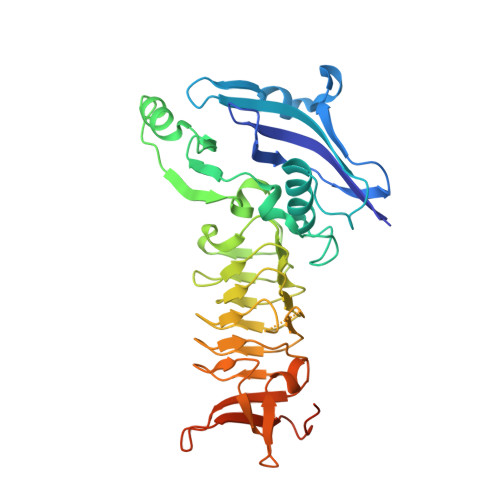
3FSY

Structure of tetrahydrodipicolinate N-succinyltransferase (Rv1201c;DapD) in complex with succinyl-CoA from Mycobacterium tuberculosis


Experimental Data Snapshot

  • Method: X-RAY DIFFRACTION
  • Resolution: 1.97 Å
  • R-Value Free: 0.200 
  • R-Value Work: 0.161 
  • R-Value Observed: 0.161 

wwPDB Validation   3D Report Full Report


Ligand Structure Quality Assessment 


This is version 1.3 of the entry. See complete history


Literature

The three-dimensional Structure of a mycobacterial DapD provides insights into DapD diversity and reveals unexpected particulars about the enzymatic mechanism.

Schuldt, L.Weyand, S.Kefala, G.Weiss, M.S.

(2009) J Mol Biol 389: 863-879

  • DOI: https://doi.org/10.1016/j.jmb.2009.04.046
  • Primary Citation of Related Structures:  
    3FSX, 3FSY

  • PubMed Abstract: 

    The enzyme tetrahydrodipicolinate N-succinyltransferase (DapD) is part of the L-lysine biosynthetic pathway. This pathway is crucial for the survival of the pathogen Mycobacterium tuberculosis (Mtb) and, consequently, the enzymes of the pathway are potential drug targets. We report here the crystal structures of Mtb-DapD and of Mtb-DapD in complex with the co-factor succinyl-CoA (SCoA) at 2.15 A and 1.97 A resolution, respectively. Each subunit of the trimeric enzyme consists of three domains, of which the second, a left-handed, parallel beta-helix (LbetaH domain), is the common structural motif of enzymes belonging to the hexapeptide repeat superfamily. The trimeric quaternary structure is stabilized by Mg(2+) and Na(+) located on the 3-fold axis. The binary complex of Mtb-DapD and SCoA reveals the binding mode(s) of the co-factor and a possible covalent reaction intermediate. The N-terminal domain of Mtb-DapD exhibits a unique architecture, including an interior water-filled channel, which allows access to a magnesium ion located at the 3-fold symmetry axis.


  • Organizational Affiliation

    EMBL Hamburg Outstation, Germany.


Macromolecules
Find similar proteins by:  (by identity cutoff)  |  3D Structure
Entity ID: 1
MoleculeChains Sequence LengthOrganismDetailsImage
Tetrahydrodipicolinate N-succinyltransferase
A, B, C, D, E
332Mycobacterium tuberculosis H37RvMutation(s): 0 
Gene Names: MT1239Rv1201c
EC: 2.3.1.117
UniProt
Find proteins for P9WP21 (Mycobacterium tuberculosis (strain ATCC 25618 / H37Rv))
Explore P9WP21 
Go to UniProtKB:  P9WP21
Entity Groups  
Sequence Clusters30% Identity50% Identity70% Identity90% Identity95% Identity100% Identity
UniProt GroupP9WP21
Sequence Annotations
Expand
  • Reference Sequence
Small Molecules
Ligands 5 Unique
IDChains Name / Formula / InChI Key2D Diagram3D Interactions
SCA
Query on SCA

Download Ideal Coordinates CCD File 
F [auth A],
H [auth A],
N [auth B],
Y [auth E]
SUCCINYL-COENZYME A
C25 H40 N7 O19 P3 S
VNOYUJKHFWYWIR-ITIYDSSPSA-N
MPD
Query on MPD

Download Ideal Coordinates CCD File 
G [auth A],
P [auth B],
U [auth D]
(4S)-2-METHYL-2,4-PENTANEDIOL
C6 H14 O2
SVTBMSDMJJWYQN-YFKPBYRVSA-N
ACY
Query on ACY

Download Ideal Coordinates CCD File 
O [auth B],
S [auth C],
T [auth D]
ACETIC ACID
C2 H4 O2
QTBSBXVTEAMEQO-UHFFFAOYSA-N
MG
Query on MG

Download Ideal Coordinates CCD File 
AA [auth E]
BA [auth E]
I [auth A]
J [auth A]
K [auth A]
AA [auth E],
BA [auth E],
I [auth A],
J [auth A],
K [auth A],
L [auth A],
Q [auth B],
V [auth D],
W [auth D],
Z [auth E]
MAGNESIUM ION
Mg
JLVVSXFLKOJNIY-UHFFFAOYSA-N
NA
Query on NA

Download Ideal Coordinates CCD File 
CA [auth E],
M [auth A],
R [auth B],
X [auth D]
SODIUM ION
Na
FKNQFGJONOIPTF-UHFFFAOYSA-N
Experimental Data & Validation

Experimental Data

  • Method: X-RAY DIFFRACTION
  • Resolution: 1.97 Å
  • R-Value Free: 0.200 
  • R-Value Work: 0.161 
  • R-Value Observed: 0.161 
  • Space Group: I 21 3
Unit Cell:
Length ( Å )Angle ( ˚ )
a = 216.96α = 90
b = 216.96β = 90
c = 216.96γ = 90
Software Package:
Software NamePurpose
HKL-2000data collection
REFMACrefinement
HKL-2000data reduction
SCALEPACKdata scaling

Structure Validation

View Full Validation Report


Ligand Structure Quality Assessment 


Entry History 

Deposition Data

Revision History  (Full details and data files)

  • Version 1.0: 2009-06-23
    Type: Initial release
  • Version 1.1: 2011-07-13
    Changes: Advisory, Refinement description, Version format compliance
  • Version 1.2: 2017-11-01
    Changes: Refinement description
  • Version 1.3: 2024-02-21
    Changes: Data collection, Database references, Derived calculations