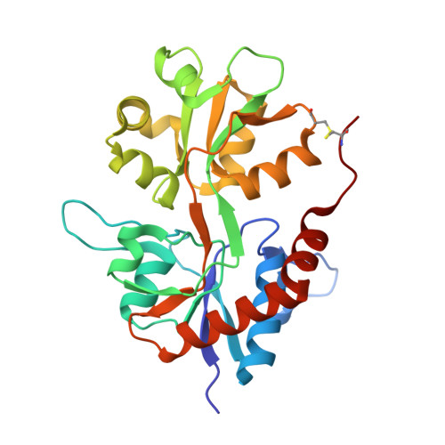
3G3I

Crystal structure of the GluR6 ligand binding domain dimer I442H K494E I749L Q753K mutant with glutamate and NaCl at 1.37 Angstrom resolution


Experimental Data Snapshot

  • Method: X-RAY DIFFRACTION
  • Resolution: 1.37 Å
  • R-Value Free: 0.176 
  • R-Value Work: 0.149 
  • R-Value Observed: 0.151 

wwPDB Validation   3D Report Full Report


This is version 1.5 of the entry. See complete history


Literature

Stability of ligand-binding domain dimer assembly controls kainate receptor desensitization.

Chaudhry, C.Weston, M.C.Schuck, P.Rosenmund, C.Mayer, M.L.

(2009) EMBO J 28: 1518-1530

  • DOI: https://doi.org/10.1038/emboj.2009.86
  • Primary Citation of Related Structures:  
    3G3F, 3G3G, 3G3H, 3G3I, 3G3J, 3G3K

  • PubMed Abstract: 

    AMPA and kainate receptors mediate fast synaptic transmission. AMPA receptor ligand-binding domains form dimers, which are key functional units controlling ion-channel activation and desensitization. Dimer stability is inversely related to the rate and extent of desensitization. Kainate and AMPA receptors share common structural elements, but functional measurements suggest that subunit assembly and gating differs between these subtypes. To investigate this, we constructed a library of GluR6 kainate receptor mutants and directly measured changes in kainate receptor dimer stability by analytical ultracentrifugation, which, combined with electrophysiological experiments, revealed an inverse correlation between dimer stability and the rate of desensitization. We solved crystal structures for a series of five GluR6 mutants, to understand the molecular mechanisms for dimer stabilization. We demonstrate that the desensitized state of kainate receptors acts as a deep energy well offsetting the stabilizing effects of dimer interface mutants, and that the deactivation of kainate receptor responses is dominated by entry into desensitized states. Our results show how neurotransmitter receptors with similar structures and gating mechanisms can exhibit strikingly different functional properties.


  • Organizational Affiliation

    Laboratory of Cellular and Molecular Neurophysiology, Porter Neuroscience Research Center, NICHD, NIH, DHHS, Bethesda, MD, USA.


Macromolecules
Find similar proteins by:  (by identity cutoff)  |  3D Structure
Entity ID: 1
MoleculeChains Sequence LengthOrganismDetailsImage
Glutamate receptor, ionotropic kainate 2
A, B
259Rattus norvegicusMutation(s): 4 
Gene Names: GriK2
UniProt
Find proteins for P42260 (Rattus norvegicus)
Explore P42260 
Go to UniProtKB:  P42260
Entity Groups  
Sequence Clusters30% Identity50% Identity70% Identity90% Identity95% Identity100% Identity
UniProt GroupP42260
Sequence Annotations
Expand
  • Reference Sequence
Experimental Data & Validation

Experimental Data

  • Method: X-RAY DIFFRACTION
  • Resolution: 1.37 Å
  • R-Value Free: 0.176 
  • R-Value Work: 0.149 
  • R-Value Observed: 0.151 
  • Space Group: P 1 21 1
Unit Cell:
Length ( Å )Angle ( ˚ )
a = 51.087α = 90
b = 114.3β = 114.99
c = 52.319γ = 90
Software Package:
Software NamePurpose
HKL-2000data collection
PHENIXrefinement
HKL-2000data reduction
HKL-2000data scaling

Structure Validation

View Full Validation Report


Entry History 

Deposition Data

Revision History  (Full details and data files)

  • Version 1.0: 2009-06-02
    Type: Initial release
  • Version 1.1: 2011-07-13
    Changes: Version format compliance
  • Version 1.2: 2017-07-26
    Changes: Source and taxonomy
  • Version 1.3: 2017-11-01
    Changes: Refinement description
  • Version 1.4: 2021-10-20
    Changes: Database references, Derived calculations
  • Version 1.5: 2024-11-20
    Changes: Data collection, Structure summary