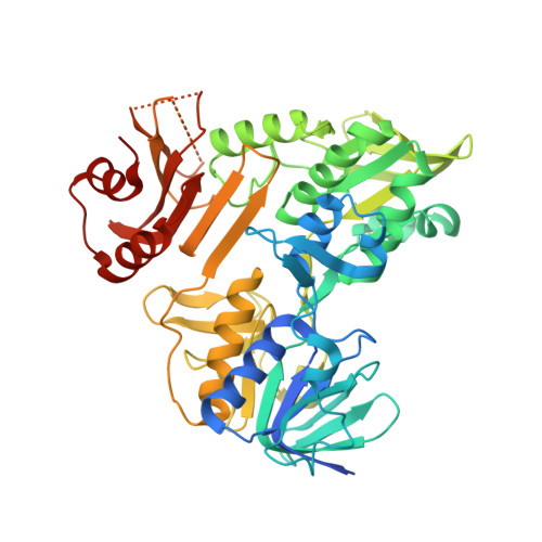
Redox-linked conformational dynamics in apoptosis-inducing factor
Sevrioukova, I.F.(2009) J Mol Biol 390: 924-938
- PubMed: 19447115
- DOI: https://doi.org/10.1016/j.jmb.2009.05.013
- Primary Citation of Related Structures:
3GD3, 3GD4 - PubMed Abstract:
Apoptosis-inducing factor (AIF) is a bifunctional mitochondrial flavoprotein critical for energy metabolism and induction of caspase-independent apoptosis, whose exact role in normal mitochondria remains unknown. Upon reduction with NADH, AIF undergoes dimerization and forms tight, long-lived FADH(2)-NAD charge-transfer complexes (CTC) that are proposed to be functionally important. To obtain a deeper insight into structure/function relations and redox mechanism of this vitally important protein, we determined the X-ray structures of oxidized and NADH-reduced forms of naturally folded recombinant murine AIF. Our structures reveal that CTC with the pyridine nucleotide is stabilized by (i) pi-stacking interactions between coplanar nicotinamide, isoalloxazine, and Phe309 rings; (ii) rearrangement of multiple aromatic residues in the C-terminal domain, likely serving as an electron delocalization site; and (iii) an extensive hydrogen-bonding network involving His453, a key residue that undergoes a conformational switch to directly interact with and optimally orient the nicotinamide for charge transfer. Via the His453-containing peptide, redox changes in the active site are transmitted to the surface, promoting AIF dimerization and restricting access to a primary nuclear localization signal through which the apoptogenic form is transported to the nucleus. Structural findings agree with biochemical data and support the hypothesis that both normal and apoptogenic functions of AIF are controlled by NADH.
Organizational Affiliation:
Department of Molecular Biology and Biochemistry, University of California, Irvine, 92697-3900, USA. [email protected]


