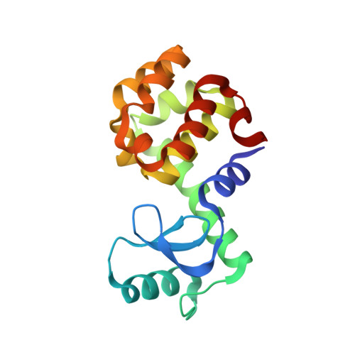
Predicting ligand binding affinity with alchemical free energy methods in a polar model binding site.
Boyce, S.E., Mobley, D.L., Rocklin, G.J., Graves, A.P., Dill, K.A., Shoichet, B.K.(2009) J Mol Biol 394: 747-763
- PubMed: 19782087
- DOI: https://doi.org/10.1016/j.jmb.2009.09.049
- Primary Citation of Related Structures:
3HT6, 3HT7, 3HT8, 3HT9, 3HTB, 3HTD, 3HTF, 3HTG, 3HU8, 3HU9, 3HUA, 3HUK, 3HUQ - PubMed Abstract:
We present a combined experimental and modeling study of organic ligand molecules binding to a slightly polar engineered cavity site in T4 lysozyme (L99A/M102Q). For modeling, we computed alchemical absolute binding free energies. These were blind tests performed prospectively on 13 diverse, previously untested candidate ligand molecules. We predicted that eight compounds would bind to the cavity and five would not; 11 of 13 predictions were correct at this level. The RMS error to the measurable absolute binding energies was 1.8 kcal/mol. In addition, we computed "relative" binding free energies for six phenol derivatives starting from two known ligands: phenol and catechol. The average RMS error in the relative free energy prediction was 2.5 kcal/mol (phenol) and 1.1 kcal/mol (catechol). To understand these results at atomic resolution, we obtained x-ray co-complex structures for nine of the diverse ligands and for all six phenol analogs. The average RMSD of the predicted pose to the experiment was 2.0 A (diverse set), 1.8 A (phenol-derived predictions), and 1.2 A (catechol-derived predictions). We found that predicting accurate affinities and rank-orderings required near-native starting orientations of the ligand in the binding site. Unanticipated binding modes, multiple ligand binding, and protein conformational change all proved challenging for the free energy methods. We believe that these results can help guide future improvements in physics-based absolute binding free energy methods.
Organizational Affiliation:
Graduate Group in Chemistry and Chemical Biology, University of California-San Francisco, San Francisco, CA 94158-2518, USA.


