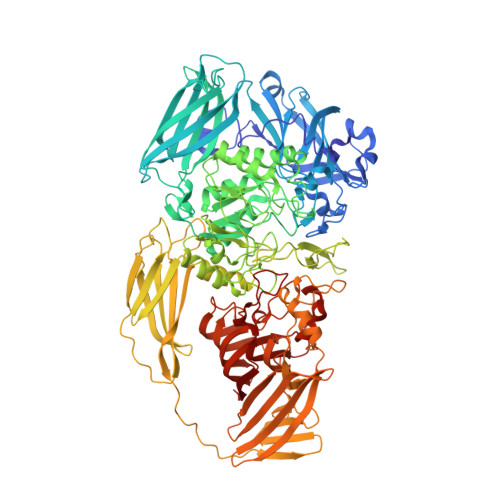
3IAP

E. coli (lacZ) beta-galactosidase (E416Q)


Experimental Data Snapshot

  • Method: X-RAY DIFFRACTION
  • Resolution: 2.00 Å
  • R-Value Free: 0.220 
  • R-Value Work: 0.181 
  • R-Value Observed: 0.181 

Starting Model: experimental
View more details

wwPDB Validation   3D Report Full Report


This is version 1.5 of the entry. See complete history


Literature

Studies of Glu-416 variants of beta-galactosidase (E. coli) show that the active site Mg(2+) is not important for structure and indicate that the main role of Mg (2+) is to mediate optimization of active site chemistry

Lo, S.Dugdale, M.L.Jeerh, N.Ku, T.Roth, N.J.Huber, R.E.

(2010) Protein J 29: 26-31

  • DOI: https://doi.org/10.1007/s10930-009-9216-x
  • Primary Citation of Related Structures:  
    3IAP, 3IAQ

  • PubMed Abstract: 

    Variants of beta-galactosidase with Valine and with Glutamine replacing Glutamate-416 did not have a Mg(2+) bound at the active site even at high Mg(2+) concentrations (200 mM). They had low catalytic activity and the pH profiles were very different from those of the native enzyme. In addition, substrates, substrate analogs, transition state analogs and galactose bound very poorly. However, the orientation and conformation of the Mg(2+) ligands (residues 416, 418, and 461) as well as the B-factors of these three side chains did not change significantly. The structures, conformations and B-factors of other active site residues were also essentially unchanged. These studies show that the active site Mg(2+) is not necessary for structure and is, therefore, mainly important for modulating the chemistry and mediating the interactions between the active site components.


  • Organizational Affiliation

    Department of Biological Sciences, University of Calgary, Calgary, AB, T2N 1N4, Canada.


Macromolecules
Find similar proteins by:  (by identity cutoff)  |  3D Structure
Entity ID: 1
MoleculeChains Sequence LengthOrganismDetailsImage
Beta-galactosidase
A, B, C, D
1,023Escherichia coli K-12Mutation(s): 1 
Gene Names: lacZ
EC: 3.2.1.23
UniProt
Find proteins for P00722 (Escherichia coli (strain K12))
Explore P00722 
Go to UniProtKB:  P00722
Entity Groups  
Sequence Clusters30% Identity50% Identity70% Identity90% Identity95% Identity100% Identity
UniProt GroupP00722
Sequence Annotations
Expand
  • Reference Sequence
Small Molecules
Ligands 4 Unique
IDChains Name / Formula / InChI Key2D Diagram3D Interactions
BTB
Query on BTB

Download Ideal Coordinates CCD File 
K [auth A],
MC [auth C],
XA [auth B],
YD [auth D]
2-[BIS-(2-HYDROXY-ETHYL)-AMINO]-2-HYDROXYMETHYL-PROPANE-1,3-DIOL
C8 H19 N O5
OWMVSZAMULFTJU-UHFFFAOYSA-N
DMS
Query on DMS

Download Ideal Coordinates CCD File 
AA [auth A]
AB [auth B]
AC [auth B]
AD [auth C]
AE [auth D]
AA [auth A],
AB [auth B],
AC [auth B],
AD [auth C],
AE [auth D],
AF [auth D],
BA [auth A],
BB [auth B],
BC [auth B],
BD [auth C],
BE [auth D],
CA [auth A],
CB [auth B],
CC [auth B],
CD [auth C],
CE [auth D],
DA [auth A],
DB [auth B],
DC [auth B],
DD [auth C],
DE [auth D],
EA [auth A],
EB [auth B],
EC [auth B],
ED [auth C],
EE [auth D],
FA [auth A],
FB [auth B],
FC [auth C],
FD [auth C],
FE [auth D],
GA [auth A],
GB [auth B],
GD [auth C],
GE [auth D],
HA [auth A],
HB [auth B],
HD [auth C],
HE [auth D],
IA [auth A],
IB [auth B],
ID [auth C],
IE [auth D],
JA [auth A],
JB [auth B],
JD [auth C],
JE [auth D],
KA [auth A],
KB [auth B],
KD [auth C],
KE [auth D],
L [auth A],
LA [auth A],
LB [auth B],
LD [auth C],
LE [auth D],
M [auth A],
MA [auth A],
MB [auth B],
MD [auth C],
ME [auth D],
N [auth A],
NA [auth A],
NB [auth B],
NC [auth C],
ND [auth C],
NE [auth D],
O [auth A],
OA [auth A],
OB [auth B],
OC [auth C],
OD [auth C],
OE [auth D],
P [auth A],
PA [auth A],
PB [auth B],
PC [auth C],
PD [auth C],
PE [auth D],
Q [auth A],
QA [auth A],
QB [auth B],
QC [auth C],
QD [auth C],
QE [auth D],
R [auth A],
RB [auth B],
RC [auth C],
RD [auth C],
RE [auth D],
S [auth A],
SB [auth B],
SC [auth C],
SE [auth D],
T [auth A],
TB [auth B],
TC [auth C],
TE [auth D],
U [auth A],
UB [auth B],
UC [auth C],
UE [auth D],
V [auth A],
VB [auth B],
VC [auth C],
VE [auth D],
W [auth A],
WB [auth B],
WC [auth C],
WE [auth D],
X [auth A],
XB [auth B],
XC [auth C],
XE [auth D],
Y [auth A],
YA [auth B],
YB [auth B],
YC [auth C],
YE [auth D],
Z [auth A],
ZA [auth B],
ZB [auth B],
ZC [auth C],
ZD [auth D],
ZE [auth D]
DIMETHYL SULFOXIDE
C2 H6 O S
IAZDPXIOMUYVGZ-UHFFFAOYSA-N
MG
Query on MG

Download Ideal Coordinates CCD File 
E [auth A]
F [auth A]
GC [auth C]
HC [auth C]
RA [auth B]
E [auth A],
F [auth A],
GC [auth C],
HC [auth C],
RA [auth B],
SA [auth B],
SD [auth D],
TD [auth D]
MAGNESIUM ION
Mg
JLVVSXFLKOJNIY-UHFFFAOYSA-N
NA
Query on NA

Download Ideal Coordinates CCD File 
G [auth A]
H [auth A]
I [auth A]
IC [auth C]
J [auth A]
G [auth A],
H [auth A],
I [auth A],
IC [auth C],
J [auth A],
JC [auth C],
KC [auth C],
LC [auth C],
TA [auth B],
UA [auth B],
UD [auth D],
VA [auth B],
VD [auth D],
WA [auth B],
WD [auth D],
XD [auth D]
SODIUM ION
Na
FKNQFGJONOIPTF-UHFFFAOYSA-N
Experimental Data & Validation

Experimental Data

  • Method: X-RAY DIFFRACTION
  • Resolution: 2.00 Å
  • R-Value Free: 0.220 
  • R-Value Work: 0.181 
  • R-Value Observed: 0.181 
  • Space Group: P 21 21 21
Unit Cell:
Length ( Å )Angle ( ˚ )
a = 151.23α = 90
b = 162.34β = 90
c = 202.06γ = 90
Software Package:
Software NamePurpose
REFMACrefinement
CNSrefinement
ADSCdata collection
MOSFLMdata reduction
SCALAdata scaling
CNSphasing

Structure Validation

View Full Validation Report


Entry History 

Deposition Data

Revision History  (Full details and data files)

  • Version 1.0: 2009-12-29
    Type: Initial release
  • Version 1.1: 2011-07-13
    Changes: Version format compliance
  • Version 1.2: 2017-11-29
    Changes: Database references
  • Version 1.3: 2019-07-24
    Changes: Data collection, Refinement description
  • Version 1.4: 2021-10-13
    Changes: Database references, Derived calculations
  • Version 1.5: 2023-09-06
    Changes: Data collection, Refinement description