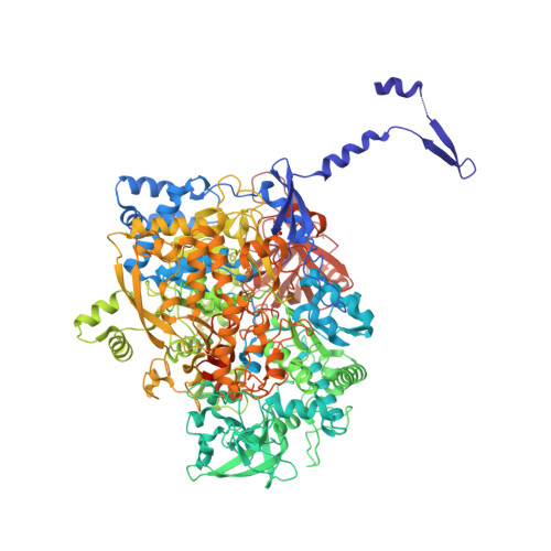
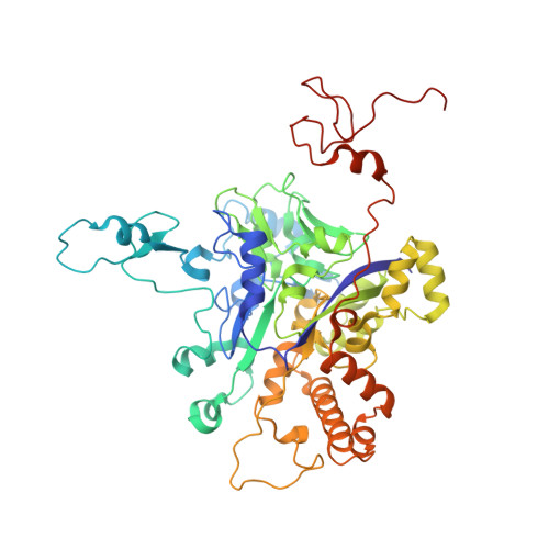
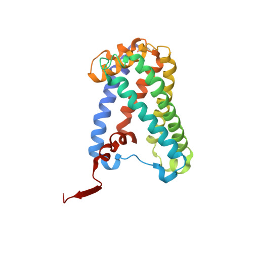
3IR6

Crystal structure of NarGHI mutant NarG-H49S


Experimental Data Snapshot

  • Method: X-RAY DIFFRACTION
  • Resolution: 2.80 Å
  • R-Value Free: 0.249 
  • R-Value Work: 0.188 

wwPDB Validation   3D Report Full Report


Ligand Structure Quality Assessment 


This is version 1.3 of the entry. See complete history


Literature

Protein crystallography reveals a role for the FS0 cluster of Escherichia coli nitrate reductase A (NarGHI) in enzyme maturation.

Rothery, R.A.Bertero, M.G.Spreter, T.Bouromand, N.Strynadka, N.C.Weiner, J.H.

(2010) J Biol Chem 285: 8801-8807

  • DOI: https://doi.org/10.1074/jbc.M109.066027
  • Primary Citation of Related Structures:  
    3IR5, 3IR6, 3IR7

  • PubMed Abstract: 

    We have used site-directed mutagenesis, EPR spectroscopy, redox potentiometry, and protein crystallography to monitor assembly of the FS0 [4Fe-4S] cluster and molybdo-bis(pyranopterin guanine dinucleotide) cofactor (Mo-bisPGD) of the Escherichia coli nitrate reductase A (NarGHI) catalytic subunit (NarG). Cys and Ser mutants of NarG-His(49) both lack catalytic activity, with only the former assembling FS0 and Mo-bisPGD. Importantly, both prosthetic groups are absent in the NarG-H49S mutant. EPR spectroscopy of the Cys mutant reveals that the E(m) value of the FS0 cluster is decreased by at least 500 mV, preventing its participation in electron transfer to the Mo-bisPGD cofactor. To demonstrate that decreasing the FS0 cluster E(m) results in decreased enzyme activity, we mutated a critical Arg residue (NarG-Arg(94)) in the vicinity of FS0 to a Ser residue. In this case, the E(m) of FS0 is decreased by 115 mV, with a concomitant decrease in enzyme turnover to approximately 30% of the wild type. Analysis of the structure of the NarG-H49S mutant reveals two important aspects of NarGHI maturation: (i) apomolybdo-NarGHI is able to bind GDP moieties at their respective P and Q sites in the absence of the Mo-bisPGD cofactor, and (ii) a critical segment of residues in NarG, (49)HGVNCTG(55), must be correctly positioned to ensure holoenzyme maturation.


  • Organizational Affiliation

    Department of Biochemistry, University of Alberta, Edmonton, Alberta T6G 2H7, Canada.


Macromolecules
Find similar proteins by:  (by identity cutoff)  |  3D Structure
Entity ID: 1
MoleculeChains Sequence LengthOrganismDetailsImage
Respiratory nitrate reductase 1 alpha chain1,247Escherichia coli K-12Mutation(s): 1 
Gene Names: narGbisDnarCb1224JW1215
EC: 1.7.99.4 (PDB Primary Data), 1.7.5.1 (UniProt)
Membrane Entity: Yes 
UniProt
Find proteins for P09152 (Escherichia coli (strain K12))
Explore P09152 
Go to UniProtKB:  P09152
Entity Groups  
Sequence Clusters30% Identity50% Identity70% Identity90% Identity95% Identity100% Identity
UniProt GroupP09152
Sequence Annotations
Expand
  • Reference Sequence
Find similar proteins by:  (by identity cutoff)  |  3D Structure
Entity ID: 2
MoleculeChains Sequence LengthOrganismDetailsImage
Respiratory nitrate reductase 1 beta chain512Escherichia coli K-12Mutation(s): 0 
Gene Names: narHb1225JW1216
EC: 1.7.99.4 (PDB Primary Data), 1.7.5.1 (UniProt)
Membrane Entity: Yes 
UniProt
Find proteins for P11349 (Escherichia coli (strain K12))
Explore P11349 
Go to UniProtKB:  P11349
Entity Groups  
Sequence Clusters30% Identity50% Identity70% Identity90% Identity95% Identity100% Identity
UniProt GroupP11349
Sequence Annotations
Expand
  • Reference Sequence
Find similar proteins by:  (by identity cutoff)  |  3D Structure
Entity ID: 3
MoleculeChains Sequence LengthOrganismDetailsImage
Respiratory nitrate reductase 1 gamma chain225Escherichia coli K-12Mutation(s): 0 
Gene Names: narIchlIb1227JW1218
EC: 1.7.99.4 (PDB Primary Data), 1.7.5.1 (UniProt)
Membrane Entity: Yes 
UniProt
Find proteins for P11350 (Escherichia coli (strain K12))
Explore P11350 
Go to UniProtKB:  P11350
Entity Groups  
Sequence Clusters30% Identity50% Identity70% Identity90% Identity95% Identity100% Identity
UniProt GroupP11350
Sequence Annotations
Expand
  • Reference Sequence
Small Molecules
Ligands 5 Unique
IDChains Name / Formula / InChI Key2D Diagram3D Interactions
HEM
Query on HEM

Download Ideal Coordinates CCD File 
K [auth C],
L [auth C]
PROTOPORPHYRIN IX CONTAINING FE
C34 H32 Fe N4 O4
KABFMIBPWCXCRK-RGGAHWMASA-L
AGA
Query on AGA

Download Ideal Coordinates CCD File 
F [auth A](1S)-2-{[{[(2S)-2,3-DIHYDROXYPROPYL]OXY}(HYDROXY)PHOSPHORYL]OXY}-1-[(PENTANOYLOXY)METHYL]ETHYL OCTANOATE
C19 H36 O10 P
UQSXQYRZHMGKIE-DLBZAZTESA-M
GDP
Query on GDP

Download Ideal Coordinates CCD File 
D [auth A],
E [auth A]
GUANOSINE-5'-DIPHOSPHATE
C10 H15 N5 O11 P2
QGWNDRXFNXRZMB-UUOKFMHZSA-N
SF4
Query on SF4

Download Ideal Coordinates CCD File 
G [auth B],
H [auth B],
I [auth B]
IRON/SULFUR CLUSTER
Fe4 S4
LJBDFODJNLIPKO-UHFFFAOYSA-N
F3S
Query on F3S

Download Ideal Coordinates CCD File 
J [auth B]FE3-S4 CLUSTER
Fe3 S4
FCXHZBQOKRZXKS-UHFFFAOYSA-N
Modified Residues  1 Unique
IDChains TypeFormula2D DiagramParent
FME
Query on FME
C
L-PEPTIDE LINKINGC6 H11 N O3 SMET
Experimental Data & Validation

Experimental Data

  • Method: X-RAY DIFFRACTION
  • Resolution: 2.80 Å
  • R-Value Free: 0.249 
  • R-Value Work: 0.188 
  • Space Group: C 2 2 21
Unit Cell:
Length ( Å )Angle ( ˚ )
a = 154.142α = 90
b = 239.825β = 90
c = 139.873γ = 90
Software Package:
Software NamePurpose
DENZOdata reduction
SCALEPACKdata scaling
CNSrefinement
PDB_EXTRACTdata extraction
HKL-2000data reduction
HKL-2000data scaling

Structure Validation

View Full Validation Report


Ligand Structure Quality Assessment 


Entry History 

Deposition Data

Revision History  (Full details and data files)

  • Version 1.0: 2010-01-05
    Type: Initial release
  • Version 1.1: 2011-07-13
    Changes: Version format compliance
  • Version 1.2: 2017-11-01
    Changes: Refinement description
  • Version 1.3: 2021-10-13
    Changes: Database references, Derived calculations