3KBS

Room Temperature X-ray structure of D-Xylose Isomerase in complex with 2Cd(2+) co-factors


Experimental Data Snapshot

  • Method: X-RAY DIFFRACTION
  • Resolution: 1.80 Å
  • R-Value Free: 0.284 
  • R-Value Observed: 0.218 

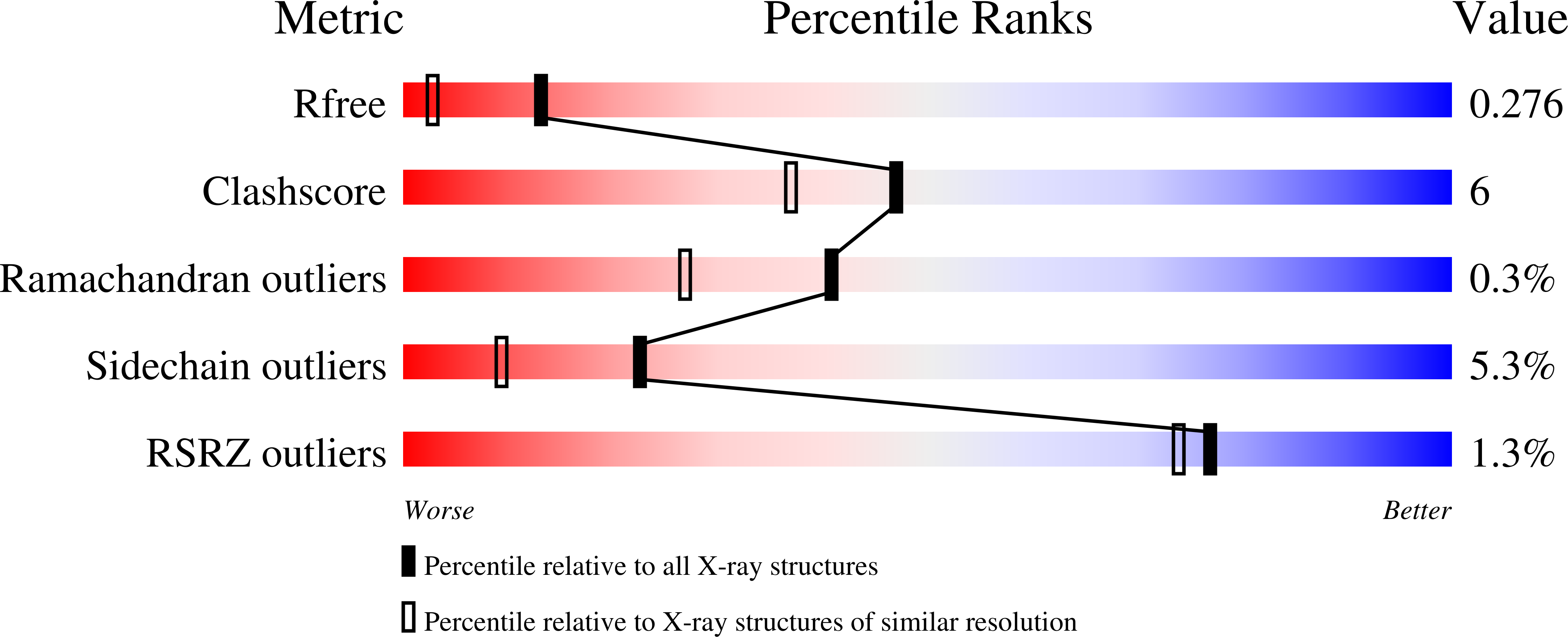
wwPDB Validation   3D Report Full Report


This is version 1.3 of the entry. See complete history


Literature

Metal ion roles and the movement of hydrogen during reaction catalyzed by D-xylose isomerase: a joint x-ray and neutron diffraction study.

Kovalevsky, A.Y.Hanson, L.Fisher, S.Z.Mustyakimov, M.Mason, S.A.Forsyth, V.T.Blakeley, M.P.Keen, D.A.Wagner, T.Carrell, H.L.Katz, A.K.Glusker, J.P.Langan, P.

(2010) Structure 18: 688-699

  • DOI: https://doi.org/10.1016/j.str.2010.03.011
  • Primary Citation of Related Structures:  
    3KBM, 3KBN, 3KBS, 3KBV, 3KBW, 3KCL, 3KCO

  • PubMed Abstract: 

    Conversion of aldo to keto sugars by the metalloenzyme D-xylose isomerase (XI) is a multistep reaction that involves hydrogen transfer. We have determined the structure of this enzyme by neutron diffraction in order to locate H atoms (or their isotope D). Two studies are presented, one of XI containing cadmium and cyclic D-glucose (before sugar ring opening has occurred), and the other containing nickel and linear D-glucose (after ring opening has occurred but before isomerization). Previously we reported the neutron structures of ligand-free enzyme and enzyme with bound product. The data show that His54 is doubly protonated on the ring N in all four structures. Lys289 is neutral before ring opening and gains a proton after this; the catalytic metal-bound water is deprotonated to hydroxyl during isomerization and O5 is deprotonated. These results lead to new suggestions as to how changes might take place over the course of the reaction.


  • Organizational Affiliation

    Bioscience Division, Los Alamos National Laboratory, MS M888, Los Alamos, NM 87545, USA. [email protected]


Macromolecules
Find similar proteins by:  (by identity cutoff)  |  3D Structure
Entity ID: 1
MoleculeChains Sequence LengthOrganismDetailsImage
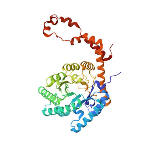
Xylose isomerase388Streptomyces rubiginosusMutation(s): 0 
Gene Names: xylA
EC: 5.3.1.5
UniProt
Find proteins for P24300 (Streptomyces rubiginosus)
Explore P24300 
Go to UniProtKB:  P24300
Entity Groups  
Sequence Clusters30% Identity50% Identity70% Identity90% Identity95% Identity100% Identity
UniProt GroupP24300
Sequence Annotations
Expand
  • Reference Sequence
Experimental Data & Validation

Experimental Data

  • Method: X-RAY DIFFRACTION
  • Resolution: 1.80 Å
  • R-Value Free: 0.284 
  • R-Value Observed: 0.218 
  • Space Group: I 2 2 2
Unit Cell:
Length ( Å )Angle ( ˚ )
a = 93.63α = 90
b = 99.54β = 90
c = 102.9γ = 90
Software Package:
Software NamePurpose
CrystalCleardata collection
SHELXmodel building
SHELXL-97refinement
CrystalCleardata reduction
CrystalCleardata scaling
SHELXphasing

Structure Validation

View Full Validation Report


Entry History 

Deposition Data

Revision History  (Full details and data files)

  • Version 1.0: 2010-06-16
    Type: Initial release
  • Version 1.1: 2011-07-13
    Changes: Version format compliance
  • Version 1.2: 2012-03-07
    Changes: Database references
  • Version 1.3: 2024-02-21
    Changes: Data collection, Database references, Derived calculations