3KK2

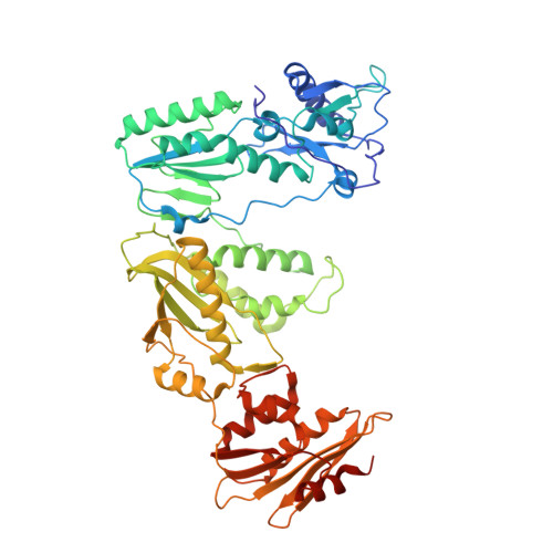
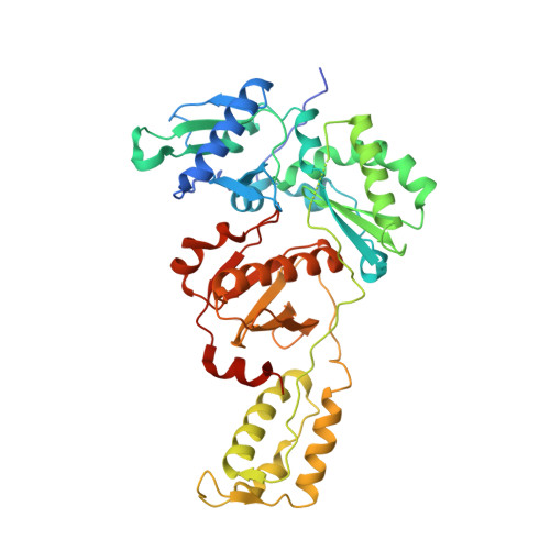
HIV-1 reverse transcriptase-DNA complex with dATP bound in the nucleotide binding site


Experimental Data Snapshot

  • Method: X-RAY DIFFRACTION
  • Resolution: 2.90 Å
  • R-Value Free: 0.259 
  • R-Value Work: 0.198 
  • R-Value Observed: 0.201 

Starting Model: experimental
View more details

wwPDB Validation   3D Report Full Report


Ligand Structure Quality Assessment 


This is version 1.3 of the entry. See complete history


Literature

Visualizing the molecular interactions of a nucleotide analog, GS-9148, with HIV-1 reverse transcriptase-DNA complex.

Lansdon, E.B.Samuel, D.Lagpacan, L.Brendza, K.M.White, K.L.Hung, M.Liu, X.Boojamra, C.G.Mackman, R.L.Cihlar, T.Ray, A.S.McGrath, M.E.Swaminathan, S.

(2010) J Mol Biol 397: 967-978

  • DOI: https://doi.org/10.1016/j.jmb.2010.02.019
  • Primary Citation of Related Structures:  
    3KJV, 3KK1, 3KK2, 3KK3

  • PubMed Abstract: 

    GS-9148 ([5-(6-amino-purin-9-yl)-4-fluoro-2,5-dihydro-furan-2-yloxymethyl]-phosphonic acid) is a dAMP (2'-deoxyadenosine monophosphate) analog that maintains its antiviral activity against drug-resistant HIV. Crystal structures for HIV-1 reverse transcriptase (RT) bound to double-stranded DNA, ternary complexes with either GS-9148-diphosphate or 2'-deoxyadenosine triphosphate (dATP), and a post-incorporation structure with GS-9148 translocated to the priming site were obtained to gain insight into the mechanism of RT inhibition. The binding of either GS-9148-diphosphate or dATP to the binary RT-DNA complex resulted in the fingers subdomain closing around the incoming substrate. This produced up to a 9 A shift in the tips of the fingers subdomain as it closed toward the palm and thumb subdomains. GS-9148-diphosphate shows a similar binding mode as dATP in the nucleotide-binding site. Residues whose mutations confer resistance to nucleotide/nucleoside RT inhibitors, such as M184, Y115, L74, and K65, show little to no shift in orientation whether GS-9148-diphosphate or dATP is bound. One difference observed in binding is the position of the central ring. The dihydrofuran ring of GS-9148-diphosphate interacts with the aromatic side chain of Y115 more than does the ribose ring of dATP, possibly picking up a favorable pi-pi interaction. The ability of GS-9148-diphosphate to mimic the active-site contacts of dATP may explain its effective inhibition of RT and maintained activity against resistance mutations. Interestingly, the 2'-fluoro moiety of GS-9148-diphosphate was found in close proximity to the Q151 side chain, potentially explaining the observed moderately reduced susceptibly to GS-9148 conferred by Q151M mutation.


  • Organizational Affiliation

    Gilead Sciences, Inc., 333 Lakeside Drive, Foster City, CA 94404, USA. [email protected]


Macromolecules

Find similar proteins by:  (by identity cutoff)  |  3D Structure
Entity ID: 1
MoleculeChains Sequence LengthOrganismDetailsImage
Reverse transcriptase p66 subunit560HIV-1 M:B_HXB2RMutation(s): 2 
Gene Names: gag-pol
EC: 2.7.7.49
UniProt
Find proteins for P04585 (Human immunodeficiency virus type 1 group M subtype B (isolate HXB2))
Explore P04585 
Go to UniProtKB:  P04585
Entity Groups  
Sequence Clusters30% Identity50% Identity70% Identity90% Identity95% Identity100% Identity
UniProt GroupP04585
Sequence Annotations
Expand
  • Reference Sequence
Find similar proteins by:  (by identity cutoff)  |  3D Structure
Entity ID: 2
MoleculeChains Sequence LengthOrganismDetailsImage
Reverse transcriptase p51 subunit452HIV-1 M:B_HXB2RMutation(s): 1 
Gene Names: gag-pol
EC: 2.7.7.49
UniProt
Find proteins for P04585 (Human immunodeficiency virus type 1 group M subtype B (isolate HXB2))
Explore P04585 
Go to UniProtKB:  P04585
Entity Groups  
Sequence Clusters30% Identity50% Identity70% Identity90% Identity95% Identity100% Identity
UniProt GroupP04585
Sequence Annotations
Expand
  • Reference Sequence

Find similar nucleic acids by:  Sequence   |   3D Structure  

Entity ID: 3
MoleculeChains LengthOrganismImage
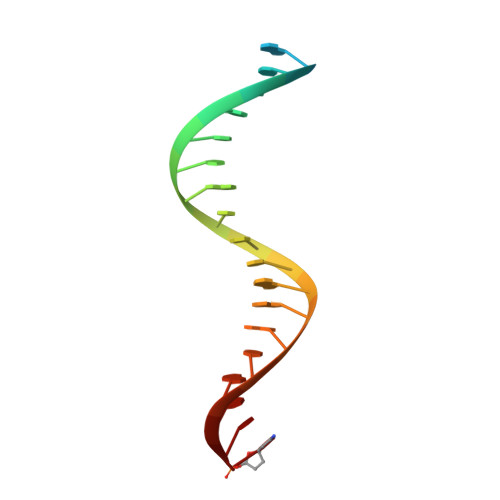
5'-D(*AP*CP*A*GP*TP*CP*CP*CP*TP*GP*TP*TP*CP*GP*GP*GP*CP*GP*CP*CP*(DOC))-3'C [auth P]21N/A
Sequence Annotations
Expand
  • Reference Sequence
Find similar nucleic acids by:  (by identity cutoff)  |  3D Structure
Entity ID: 4
MoleculeChains LengthOrganismImage
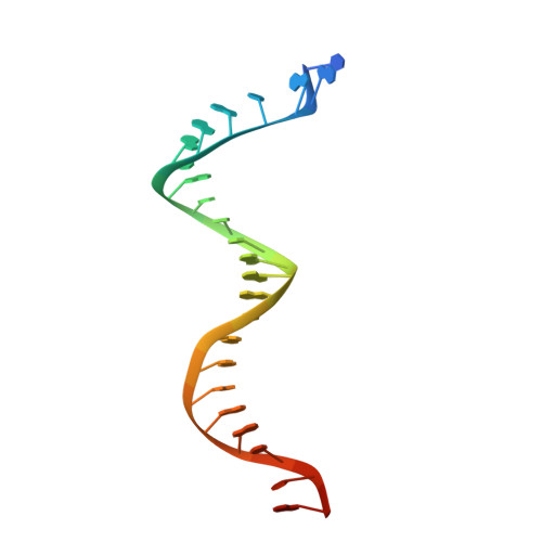
5'-D(*A*TP*GP*GP*TP*GP*GP*GP*CP*GP*CP*CP*CP*GP*AP*AP*CP*AP*GP*GP*GP*AP*CP*TP*GP*TP*G)-3'D [auth T]27N/A
Sequence Annotations
Expand
  • Reference Sequence
Experimental Data & Validation

Experimental Data

  • Method: X-RAY DIFFRACTION
  • Resolution: 2.90 Å
  • R-Value Free: 0.259 
  • R-Value Work: 0.198 
  • R-Value Observed: 0.201 
  • Space Group: C 2 2 21
Unit Cell:
Length ( Å )Angle ( ˚ )
a = 167.912α = 90
b = 168.922β = 90
c = 102.041γ = 90
Software Package:
Software NamePurpose
BOSdata collection
EPMRphasing
CNXrefinement
HKL-2000data reduction
HKL-2000data scaling

Structure Validation

View Full Validation Report


Ligand Structure Quality Assessment 


Entry History 

Deposition Data

Revision History  (Full details and data files)

  • Version 1.0: 2010-03-23
    Type: Initial release
  • Version 1.1: 2011-07-13
    Changes: Version format compliance
  • Version 1.2: 2021-10-13
    Changes: Database references, Derived calculations
  • Version 1.3: 2023-09-06
    Changes: Data collection, Refinement description