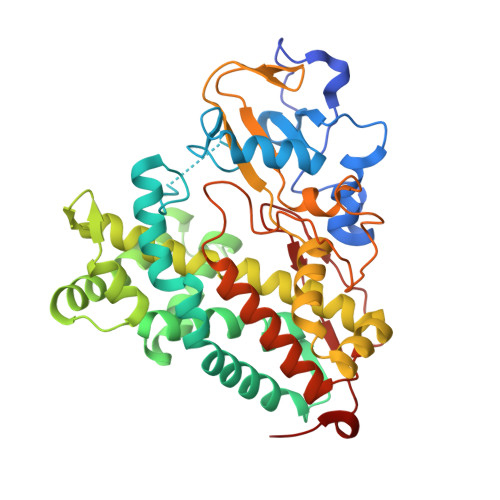
3L62

Crystal structure of substrate-free P450cam at low [K+]


Experimental Data Snapshot

  • Method: X-RAY DIFFRACTION
  • Resolution: 1.70 Å
  • R-Value Free: 0.224 
  • R-Value Work: 0.190 
  • R-Value Observed: 0.192 

Starting Model: other
View more details

wwPDB Validation   3D Report Full Report


Ligand Structure Quality Assessment 


This is version 1.4 of the entry. See complete history


Literature

P450cam visits an open conformation in the absence of substrate.

Lee, Y.T.Wilson, R.F.Rupniewski, I.Goodin, D.B.

(2010) Biochemistry 49: 3412-3419

  • DOI: https://doi.org/10.1021/bi100183g
  • Primary Citation of Related Structures:  
    3L61, 3L62, 3L63

  • PubMed Abstract: 

    P450cam from Pseudomonas putida is the best characterized member of the vast family of cytochrome P450s, and it has long been believed to have a more rigid and closed active site relative to other P450s. Here we report X-ray structures of P450cam crystallized in the absence of substrate and at high and low [K(+)]. The camphor-free structures are observed in a distinct open conformation characterized by a water-filled channel created by the retraction of the F and G helices, disorder of the B' helix, and loss of the K(+) binding site. Crystallization in the presence of K(+) alone does not alter the open conformation, while crystallization with camphor alone is sufficient for closure of the channel. Soaking crystals of the open conformation in excess camphor does not promote camphor binding or closure, suggesting resistance to conformational change by the crystal lattice. This open conformation is remarkably similar to that seen upon binding large tethered substrates, showing that it is not the result of a perturbation by the ligand. Redissolved crystals of the open conformation are observed as a mixture of P420 and P450 forms, which is converted to the P450 form upon addition of camphor and K(+). These data reveal that P450cam can dynamically visit an open conformation that allows access to the deeply buried active site without being induced by substrate or ligand.


  • Organizational Affiliation

    Department of Molecular Biology, 10550 North Torrey Pines Road, The Scripps Research Institute, La Jolla, California 92037, USA.


Macromolecules
Find similar proteins by:  (by identity cutoff)  |  3D Structure
Entity ID: 1
MoleculeChains Sequence LengthOrganismDetailsImage
Camphor 5-monooxygenase414Pseudomonas putidaMutation(s): 1 
Gene Names: camCcyp101
EC: 1.14.15.1
UniProt
Find proteins for P00183 (Pseudomonas putida)
Explore P00183 
Go to UniProtKB:  P00183
Entity Groups  
Sequence Clusters30% Identity50% Identity70% Identity90% Identity95% Identity100% Identity
UniProt GroupP00183
Sequence Annotations
Expand
  • Reference Sequence
Small Molecules
Ligands 1 Unique
IDChains Name / Formula / InChI Key2D Diagram3D Interactions
HEM
Query on HEM

Download Ideal Coordinates CCD File 
B [auth A]PROTOPORPHYRIN IX CONTAINING FE
C34 H32 Fe N4 O4
KABFMIBPWCXCRK-RGGAHWMASA-L
Experimental Data & Validation

Experimental Data

  • Method: X-RAY DIFFRACTION
  • Resolution: 1.70 Å
  • R-Value Free: 0.224 
  • R-Value Work: 0.190 
  • R-Value Observed: 0.192 
  • Space Group: P 21 21 21
Unit Cell:
Length ( Å )Angle ( ˚ )
a = 65.581α = 90
b = 73.546β = 90
c = 91.931γ = 90
Software Package:
Software NamePurpose
MOLREPphasing
REFMACrefinement
MOSFLMdata reduction
SCALAdata scaling

Structure Validation

View Full Validation Report


Ligand Structure Quality Assessment 


Entry History 

Deposition Data

Revision History  (Full details and data files)

  • Version 1.0: 2010-04-21
    Type: Initial release
  • Version 1.1: 2011-07-13
    Changes: Version format compliance
  • Version 1.2: 2021-10-13
    Changes: Database references, Derived calculations
  • Version 1.3: 2024-02-21
    Changes: Data collection
  • Version 1.4: 2024-04-03
    Changes: Refinement description