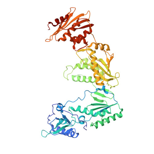
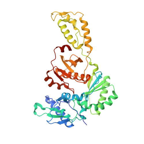
3LP1

HIV-1 reverse transcriptase with inhibitor


Experimental Data Snapshot

  • Method: X-RAY DIFFRACTION
  • Resolution: 2.23 Å
  • R-Value Free: 0.292 
  • R-Value Work: 0.268 
  • R-Value Observed: 0.269 

wwPDB Validation   3D Report Full Report


Ligand Structure Quality Assessment 


This is version 1.3 of the entry. See complete history


Literature

Structural basis for the inhibition of RNase H activity of HIV-1 reverse transcriptase by RNase H active site-directed inhibitors.

Su, H.P.Yan, Y.Prasad, G.S.Smith, R.F.Daniels, C.L.Abeywickrema, P.D.Reid, J.C.Loughran, H.M.Kornienko, M.Sharma, S.Grobler, J.A.Xu, B.Sardana, V.Allison, T.J.Williams, P.D.Darke, P.L.Hazuda, D.J.Munshi, S.

(2010) J Virol 84: 7625-7633

  • DOI: https://doi.org/10.1128/JVI.00353-10
  • Primary Citation of Related Structures:  
    3LP0, 3LP1, 3LP2, 3LP3

  • PubMed Abstract: 

    HIV/AIDS continues to be a menace to public health. Several drugs currently on the market have successfully improved the ability to manage the viral burden in infected patients. However, new drugs are needed to combat the rapid emergence of mutated forms of the virus that are resistant to existing therapies. Currently, approved drugs target three of the four major enzyme activities encoded by the virus that are critical to the HIV life cycle. Although a number of inhibitors of HIV RNase H activity have been reported, few inhibit by directly engaging the RNase H active site. Here, we describe structures of naphthyridinone-containing inhibitors bound to the RNase H active site. This class of compounds binds to the active site via two metal ions that are coordinated by catalytic site residues, D443, E478, D498, and D549. The directionality of the naphthyridinone pharmacophore is restricted by the ordering of D549 and H539 in the RNase H domain. In addition, one of the naphthyridinone-based compounds was found to bind at a second site close to the polymerase active site and non-nucleoside/nucleotide inhibitor sites in a metal-independent manner. Further characterization, using fluorescence-based thermal denaturation and a crystal structure of the isolated RNase H domain reveals that this compound can also bind the RNase H site and retains the metal-dependent binding mode of this class of molecules. These structures provide a means for structurally guided design of novel RNase H inhibitors.


  • Organizational Affiliation

    Department of Global Structural Biology, Merck Research Laboratories, West Point, Pennsylvania 19486, USA. [email protected]


Macromolecules
Find similar proteins by:  (by identity cutoff)  |  3D Structure
Entity ID: 1
MoleculeChains Sequence LengthOrganismDetailsImage
Reverse transcriptase/ribonuclease H563HIV-1 M:B_HXB2RMutation(s): 0 
Gene Names: gag-pol
EC: 2.7.7.49 (PDB Primary Data), 2.7.7.7 (PDB Primary Data), 3.1.26.4 (PDB Primary Data)
UniProt
Find proteins for P04585 (Human immunodeficiency virus type 1 group M subtype B (isolate HXB2))
Explore P04585 
Go to UniProtKB:  P04585
Entity Groups  
Sequence Clusters30% Identity50% Identity70% Identity90% Identity95% Identity100% Identity
UniProt GroupP04585
Sequence Annotations
Expand
  • Reference Sequence
Find similar proteins by:  (by identity cutoff)  |  3D Structure
Entity ID: 2
MoleculeChains Sequence LengthOrganismDetailsImage
p51 RT443HIV-1 M:B_HXB2RMutation(s): 0 
Gene Names: gag-pol
EC: 2.7.7.49 (PDB Primary Data), 2.7.7.7 (PDB Primary Data)
UniProt
Find proteins for P04585 (Human immunodeficiency virus type 1 group M subtype B (isolate HXB2))
Explore P04585 
Go to UniProtKB:  P04585
Entity Groups  
Sequence Clusters30% Identity50% Identity70% Identity90% Identity95% Identity100% Identity
UniProt GroupP04585
Sequence Annotations
Expand
  • Reference Sequence
Small Molecules
Binding Affinity Annotations 
IDSourceBinding Affinity
LP8 PDBBind:  3LP1 IC50: 120 (nM) from 1 assay(s)
Experimental Data & Validation

Experimental Data

  • Method: X-RAY DIFFRACTION
  • Resolution: 2.23 Å
  • R-Value Free: 0.292 
  • R-Value Work: 0.268 
  • R-Value Observed: 0.269 
  • Space Group: C 2 2 21
Unit Cell:
Length ( Å )Angle ( ˚ )
a = 118.784α = 90
b = 155.264β = 90
c = 155.244γ = 90
Software Package:
Software NamePurpose
DENZOdata reduction
SCALEPACKdata scaling
REFMACrefinement
PDB_EXTRACTdata extraction

Structure Validation

View Full Validation Report


Ligand Structure Quality Assessment 


Entry History 

Deposition Data

Revision History  (Full details and data files)

  • Version 1.0: 2010-06-09
    Type: Initial release
  • Version 1.1: 2011-07-13
    Changes: Version format compliance
  • Version 1.2: 2017-11-01
    Changes: Refinement description
  • Version 1.3: 2024-02-21
    Changes: Data collection, Database references, Derived calculations