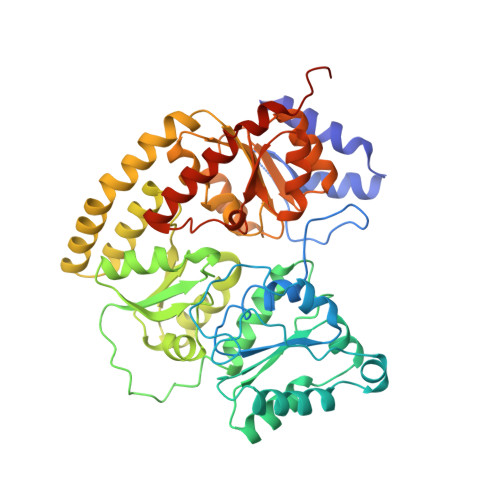
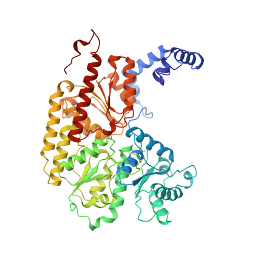
3MIN

NITROGENASE MOFE PROTEIN FROM AZOTOBACTER VINELANDII, OXIDIZED STATE


Experimental Data Snapshot

  • Method: X-RAY DIFFRACTION
  • Resolution: 2.03 Å
  • R-Value Free: 0.264 
  • R-Value Work: 0.206 
  • R-Value Observed: 0.206 

wwPDB Validation   3D Report Full Report


This is version 1.3 of the entry. See complete history


Literature

Redox-dependent structural changes in the nitrogenase P-cluster.

Peters, J.W.Stowell, M.H.Soltis, S.M.Finnegan, M.G.Johnson, M.K.Rees, D.C.

(1997) Biochemistry 36: 1181-1187

  • DOI: https://doi.org/10.1021/bi9626665
  • Primary Citation of Related Structures:  
    2MIN, 3MIN

  • PubMed Abstract: 

    The structure of the nitrogenase MoFe-protein from Azotobacter vinelandii has been refined to 2.0 A resolution in two oxidation states. EPR studies on the crystals indicate that the structures correspond to the spectroscopically assigned oxidized (P(OX)/M(OX)) and the native or dithionite-reduced (P(N)/M(N)) forms of the enzyme. Both MoFe-protein structures are essentially identical, with the exception of the P-cluster. The MoFe-protein P-cluster in each state is found to contain eight Fe and seven S atoms. Interconversion between the two redox states involves movement of two Fe atoms and an exchange of protein coordination for ligands supplied by a central S atom. In the oxidized P(OX) state, the cluster is coordinated by the protein through six cysteine ligands, Ser-beta188 O gamma, and the backbone amide of Cys-alpha88. In the native P(N) state, Ser-beta188 O gamma and the amide N of Cys-alpha88 no longer coordinate the cluster due to movement of their coordinated Fe atoms toward the central sulfur. Consequently, this central sulfur adopts a distorted octahedral environment with six surrounding Fe atoms. A previously described model of the P-cluster containing 8Fe-8S likely reflects the inappropriate modeling of a single structure to a mixture of these two P-cluster redox states. These observed redox-mediated structural changes of the P-cluster suggest a role for this cluster in coupling electron transfer and proton transfer in nitrogenase.


  • Organizational Affiliation

    Division of Chemistry and Chemical Engineering, California Institute of Technology, Pasadena 91125, USA.


Macromolecules
Find similar proteins by:  (by identity cutoff)  |  3D Structure
Entity ID: 1
MoleculeChains Sequence LengthOrganismDetailsImage
NITROGENASE MOLYBDENUM IRON PROTEIN
A, C
491Azotobacter vinelandiiMutation(s): 0 
EC: 1.18.6.1
UniProt
Find proteins for P07328 (Azotobacter vinelandii)
Explore P07328 
Go to UniProtKB:  P07328
Entity Groups  
Sequence Clusters30% Identity50% Identity70% Identity90% Identity95% Identity100% Identity
UniProt GroupP07328
Sequence Annotations
Expand
  • Reference Sequence
Find similar proteins by:  (by identity cutoff)  |  3D Structure
Entity ID: 2
MoleculeChains Sequence LengthOrganismDetailsImage
NITROGENASE MOLYBDENUM IRON PROTEIN
B, D
522Azotobacter vinelandiiMutation(s): 0 
EC: 1.18.6.1
UniProt
Find proteins for P07329 (Azotobacter vinelandii)
Explore P07329 
Go to UniProtKB:  P07329
Entity Groups  
Sequence Clusters30% Identity50% Identity70% Identity90% Identity95% Identity100% Identity
UniProt GroupP07329
Sequence Annotations
Expand
  • Reference Sequence
Small Molecules
Ligands 4 Unique
IDChains Name / Formula / InChI Key2D Diagram3D Interactions
CFM
Query on CFM

Download Ideal Coordinates CCD File 
F [auth A],
J [auth C]
FE-MO-S CLUSTER
Fe7 Mo S9
UZRXIPMKRKMLQF-UHFFFAOYSA-N
CLF
Query on CLF

Download Ideal Coordinates CCD File 
H [auth B],
L [auth D]
FE(8)-S(7) CLUSTER
Fe8 S7
JKVMXLBGZBULKV-UHFFFAOYSA-N
HCA
Query on HCA

Download Ideal Coordinates CCD File 
E [auth A],
I [auth C]
3-HYDROXY-3-CARBOXY-ADIPIC ACID
C7 H10 O7
XKJVEVRQMLKSMO-SSDOTTSWSA-N
CA
Query on CA

Download Ideal Coordinates CCD File 
G [auth B],
K [auth D]
CALCIUM ION
Ca
BHPQYMZQTOCNFJ-UHFFFAOYSA-N
Experimental Data & Validation

Experimental Data

  • Method: X-RAY DIFFRACTION
  • Resolution: 2.03 Å
  • R-Value Free: 0.264 
  • R-Value Work: 0.206 
  • R-Value Observed: 0.206 
  • Space Group: P 1 21 1
Unit Cell:
Length ( Å )Angle ( ˚ )
a = 108α = 90
b = 131.3β = 110.7
c = 81γ = 90
Software Package:
Software NamePurpose
X-PLORmodel building
X-PLORrefinement
DENZOdata reduction
SCALEPACKdata scaling
X-PLORphasing

Structure Validation

View Full Validation Report


Entry History 

Deposition Data

Revision History  (Full details and data files)

  • Version 1.0: 1997-04-01
    Type: Initial release
  • Version 1.1: 2008-03-03
    Changes: Version format compliance
  • Version 1.2: 2011-07-13
    Changes: Version format compliance
  • Version 1.3: 2024-02-21
    Changes: Data collection, Database references, Derived calculations, Other