3MVY

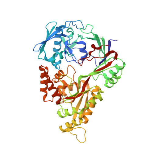
X-ray structure of the diatomic oxo-intermediate NikA/1-Int', prior hydroxylation


Experimental Data Snapshot

  • Method: X-RAY DIFFRACTION
  • Resolution: 2.50 Å
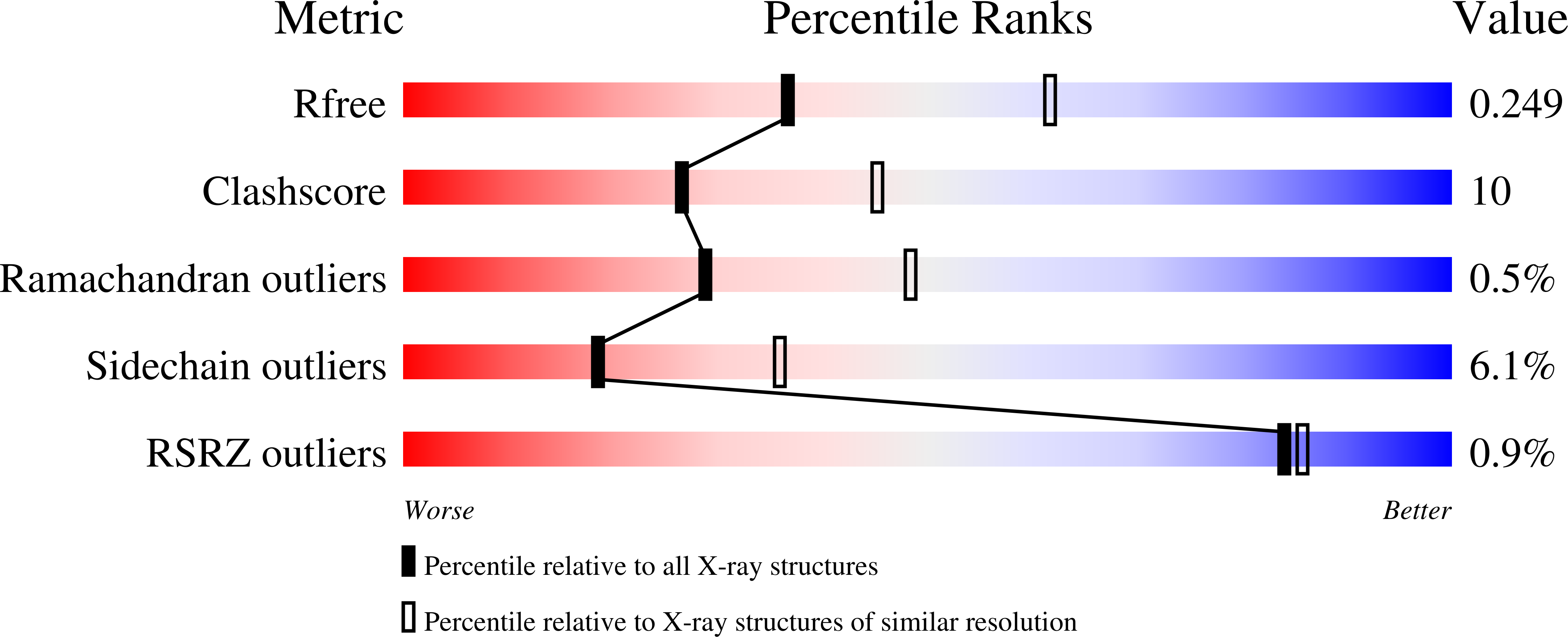
  • R-Value Free: 0.249 
  • R-Value Work: 0.173 
  • R-Value Observed: 0.176 

Starting Model: experimental
View more details

wwPDB Validation   3D Report Full Report


Ligand Structure Quality Assessment 


This is version 1.2 of the entry. See complete history


Literature

Crystallographic snapshots of the reaction of aromatic C-H with O(2) catalysed by a protein-bound iron complex

Cavazza, C.Bochot, C.Rousselot-Pailley, P.Carpentier, P.Cherrier, M.V.Martin, L.Marchi-Delapierre, C.Fontecilla-Camps, J.C.Menage, S.

(2010) Nat Chem 2: 1069-1076

  • DOI: https://doi.org/10.1038/nchem.841
  • Primary Citation of Related Structures:  
    3MVW, 3MVX, 3MVY, 3MVZ, 3MW0, 3MZ9, 3MZB

  • PubMed Abstract: 

    Chemical reactions inside single crystals are quite rare because crystallinity is difficult to retain owing to atomic rearrangements. Protein crystals in general have a high solvent content. This allows for some molecular flexibility, which makes it possible to trap reaction intermediates of enzymatic reactions without disrupting the crystal lattice. A similar approach has not yet been fully implemented in the field of inorganic chemistry. Here, we have combined model chemistry and protein X-ray crystallography to study the intramolecular aromatic dihydroxylation by an arene-containing protein-bound iron complex. The bound complex was able to activate dioxygen in the presence of a reductant, leading to the formation of catechol as the sole product. The structure determination of four of the catalytic cycle intermediates and the end product showed that the hydroxylation reaction implicates an iron peroxo, generated by reductive O(2) activation, an intermediate already observed in iron monooxygenases. This strategy also provided unexpected mechanistic details such as the rearrangement of the iron coordination sphere on metal reduction.


  • Organizational Affiliation

    Laboratoire de Cristallographie et de Cristallogenèse des Protéines, Institut de Biologie Structurale J.P. Ebel, UMR 5075 CEA, CNRS, Université Joseph Fourier, 41 rue Horowitz, 38027 Grenoble Cedex 1, France. [email protected]


Macromolecules
Find similar proteins by:  (by identity cutoff)  |  3D Structure
Entity ID: 1
MoleculeChains Sequence LengthOrganismDetailsImage
Nickel-binding periplasmic protein
A, B
502Escherichia coli K-12Mutation(s): 0 
Gene Names: nikA
EC: 3.6.3.24
UniProt
Find proteins for P33590 (Escherichia coli (strain K12))
Explore P33590 
Go to UniProtKB:  P33590
Entity Groups  
Sequence Clusters30% Identity50% Identity70% Identity90% Identity95% Identity100% Identity
UniProt GroupP33590
Sequence Annotations
Expand
  • Reference Sequence
Small Molecules
Ligands 7 Unique
IDChains Name / Formula / InChI Key2D Diagram3D Interactions
BHZ
Query on BHZ

Download Ideal Coordinates CCD File 
K [auth A],
W [auth B]
2-[2-[carboxymethyl(phenylmethyl)amino]ethyl-[(2-hydroxyphenyl)methyl]amino]ethanoic acid
C20 H24 N2 O5
JSDNXVMPVNBNSK-UHFFFAOYSA-N
SO4
Query on SO4

Download Ideal Coordinates CCD File 
F [auth A]SULFATE ION
O4 S
QAOWNCQODCNURD-UHFFFAOYSA-L
GOL
Query on GOL

Download Ideal Coordinates CCD File 
G [auth A]
H [auth A]
I [auth A]
O [auth A]
P [auth A]
G [auth A],
H [auth A],
I [auth A],
O [auth A],
P [auth A],
R [auth A],
U [auth B],
Z [auth B]
GLYCEROL
C3 H8 O3
PEDCQBHIVMGVHV-UHFFFAOYSA-N
ACT
Query on ACT

Download Ideal Coordinates CCD File 
C [auth A]
D [auth A]
E [auth A]
Q [auth A]
S [auth B]
C [auth A],
D [auth A],
E [auth A],
Q [auth A],
S [auth B],
T [auth B],
Y [auth B]
ACETATE ION
C2 H3 O2
QTBSBXVTEAMEQO-UHFFFAOYSA-M
FE
Query on FE

Download Ideal Coordinates CCD File 
J [auth A],
V [auth B]
FE (III) ION
Fe
VTLYFUHAOXGGBS-UHFFFAOYSA-N
CL
Query on CL

Download Ideal Coordinates CCD File 
M [auth A],
N [auth A]
CHLORIDE ION
Cl
VEXZGXHMUGYJMC-UHFFFAOYSA-M
PER
Query on PER

Download Ideal Coordinates CCD File 
L [auth A],
X [auth B]
PEROXIDE ION
O2
ANAIPYUSIMHBEL-UHFFFAOYSA-N
Experimental Data & Validation

Experimental Data

  • Method: X-RAY DIFFRACTION
  • Resolution: 2.50 Å
  • R-Value Free: 0.249 
  • R-Value Work: 0.173 
  • R-Value Observed: 0.176 
  • Space Group: P 21 21 21
Unit Cell:
Length ( Å )Angle ( ˚ )
a = 86.985α = 90
b = 95.172β = 90
c = 125.049γ = 90
Software Package:
Software NamePurpose
REFMACrefinement

Structure Validation

View Full Validation Report


Ligand Structure Quality Assessment 


Entry History 

Revision History  (Full details and data files)

  • Version 1.0: 2011-02-09
    Type: Initial release
  • Version 1.1: 2011-07-13
    Changes: Version format compliance
  • Version 1.2: 2023-11-01
    Changes: Data collection, Database references, Derived calculations, Refinement description