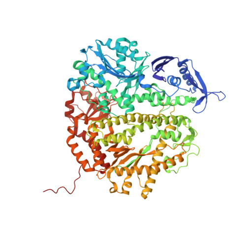
3NDK

RB69 DNA Polymerase (Y567A) Ternary Complex with dCTP Opposite dG


Experimental Data Snapshot

  • Method: X-RAY DIFFRACTION
  • Resolution: 2.00 Å
  • R-Value Free: 0.229 
  • R-Value Work: 0.187 
  • R-Value Observed: 0.189 

Starting Model: experimental
View more details

wwPDB Validation   3D Report Full Report


Ligand Structure Quality Assessment 


This is version 1.2 of the entry. See complete history


Literature

Variation in Mutation Rates Caused by RB69pol Fidelity Mutants Can Be Rationalized on the Basis of Their Kinetic Behavior and Crystal Structures.

Xia, S.Wang, M.Lee, H.R.Sinha, A.Blaha, G.Christian, T.Wang, J.Konigsberg, W.

(2011) J Mol Biol 406: 558-570

  • DOI: https://doi.org/10.1016/j.jmb.2010.12.033
  • Primary Citation of Related Structures:  
    3NDK, 3NE6, 3NGI, 3NHG

  • PubMed Abstract: 

    We have previously observed that stepwise replacement of amino acid residues in the nascent base-pair binding pocket of RB69 DNA polymerase (RB69pol) with Ala or Gly expanded the space in this pocket, resulting in a progressive increase in misincorporation. However, in vivo results with similar RB69pol nascent base-pair binding pocket mutants showed that mutation rates, as determined by the T4 phage rI forward assay and rII reversion assay, were significantly lower for the RB69pol S565G/Y567A double mutant than for the Y567A single mutant, the opposite of what we would have predicted. To investigate the reasons for this unexpected result, we have determined the pre-steady-state kinetic parameters and crystal structures of relevant ternary complexes. We found that the S565G/Y567A mutant generally had greater base selectivity than the Y567A mutant and that the kinetic parameters for dNMP insertion, excision of the 3'-terminal nucleotide residue, and primer extension beyond a mispair differed not only between these two mutants but also between the two highly mutable sequences in the T4 rI complementary strand. Comparison of the crystal structures of these two mutants with correct and incorrect incoming dNTPs provides insight into the unexpected increase in the fidelity of the S565G/Y567A double mutant. Taken together, the kinetic and structural results provide a basis for integrating and interpreting in vivo and in vitro observations.


  • Organizational Affiliation

    Department of Molecular Biophysics and Biochemistry, Yale University, 333 Cedar Street, New Haven, CT 06520, USA.


Macromolecules

Find similar proteins by:  (by identity cutoff)  |  3D Structure
Entity ID: 1
MoleculeChains Sequence LengthOrganismDetailsImage
DNA polymerase903Escherichia phage RB69Mutation(s): 3 
Gene Names: 43gp43
EC: 2.7.7.7 (PDB Primary Data), 3.1.11 (UniProt)
UniProt
Find proteins for Q38087 (Escherichia phage RB69)
Explore Q38087 
Go to UniProtKB:  Q38087
Entity Groups  
Sequence Clusters30% Identity50% Identity70% Identity90% Identity95% Identity100% Identity
UniProt GroupQ38087
Sequence Annotations
Expand
  • Reference Sequence

Find similar nucleic acids by:  Sequence   |   3D Structure  

Entity ID: 2
MoleculeChains LengthOrganismImage
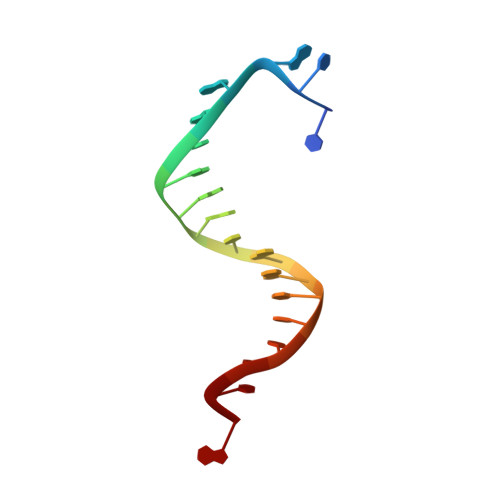
DNA (5'-D(*TP*CP*AP*GP*GP*TP*AP*AP*GP*CP*AP*GP*TP*CP*CP*GP*CP*G)-3')B [auth T]18N/A
Sequence Annotations
Expand
  • Reference Sequence

Find similar nucleic acids by:  Sequence   |   3D Structure  

Entity ID: 3
MoleculeChains LengthOrganismImage
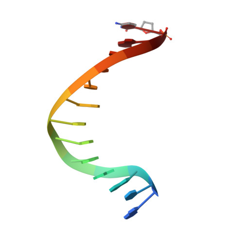
DNA (5'-D(*GP*CP*GP*GP*AP*CP*TP*GP*CP*TP*TP*AP*(DOC))-3')C [auth P]13N/A
Sequence Annotations
Expand
  • Reference Sequence
Small Molecules
Ligands 2 Unique
IDChains Name / Formula / InChI Key2D Diagram3D Interactions
DCP
Query on DCP

Download Ideal Coordinates CCD File 
D [auth A]2'-DEOXYCYTIDINE-5'-TRIPHOSPHATE
C9 H16 N3 O13 P3
RGWHQCVHVJXOKC-SHYZEUOFSA-N
CA
Query on CA

Download Ideal Coordinates CCD File 
E [auth A]
F [auth A]
G [auth A]
H [auth A]
I [auth A]
E [auth A],
F [auth A],
G [auth A],
H [auth A],
I [auth A],
J [auth A],
K [auth A]
CALCIUM ION
Ca
BHPQYMZQTOCNFJ-UHFFFAOYSA-N
Experimental Data & Validation

Experimental Data

  • Method: X-RAY DIFFRACTION
  • Resolution: 2.00 Å
  • R-Value Free: 0.229 
  • R-Value Work: 0.187 
  • R-Value Observed: 0.189 
  • Space Group: P 21 21 21
Unit Cell:
Length ( Å )Angle ( ˚ )
a = 75.006α = 90
b = 120.015β = 90
c = 130.781γ = 90
Software Package:
Software NamePurpose
HKL-2000data collection
AMoREphasing
REFMACrefinement
HKL-2000data reduction
HKL-2000data scaling

Structure Validation

View Full Validation Report


Ligand Structure Quality Assessment 


Entry History 

Deposition Data

Revision History  (Full details and data files)

  • Version 1.0: 2011-01-26
    Type: Initial release
  • Version 1.1: 2011-07-13
    Changes: Version format compliance
  • Version 1.2: 2023-09-06
    Changes: Data collection, Database references, Derived calculations, Refinement description