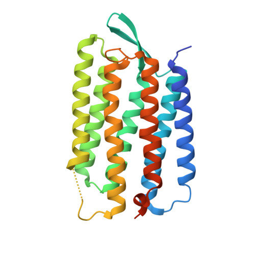
X-ray-Radiation-Induced Changes in Bacteriorhodopsin Structure.
Borshchevskiy, V.I., Round, E.S., Popov, A.N., Buldt, G., Gordeliy, V.I.(2011) J Mol Biol 409: 813-825
- PubMed: 21530535
- DOI: https://doi.org/10.1016/j.jmb.2011.04.038
- Primary Citation of Related Structures:
3NS0, 3NSB - PubMed Abstract:
Bacteriorhodopsin (bR) provides light-driven vectorial proton transport across a cell membrane. Creation of electrochemical potential at the membrane is a universal step in energy transformation in a cell. Published atomic crystallographic models of early intermediate states of bR show a significant difference between them, and conclusions about pumping mechanisms have been contradictory. Here, we present a quantitative high-resolution crystallographic study of conformational changes in bR induced by X-ray absorption. It is shown that X-ray doses that are usually accumulated during data collection for intermediate-state studies are sufficient to significantly alter the structure of the protein. X-ray-induced changes occur primarily in the active site of bR. Structural modeling showed that X-ray absorption triggers retinal isomerization accompanied by the disappearance of electron densities corresponding to the water molecule W402 bound to the Schiff base. It is demonstrated that these and other X-ray-induced changes may mimic functional conformational changes of bR leading to misinterpretation of the earlier obtained X-ray crystallographic structures of photointermediates.
Organizational Affiliation:
Laboratoire des Protéines Membranaires, Institut de Biologie Structurale J.-P. Ebel, UMR5075 CEA-CNRS-UJF, Grenoble 38027, France.


