3NTY

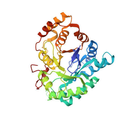
Crystal structure of AKR1C1 in complex with NADP and 5-Phenyl,3-chlorosalicylic acid


Experimental Data Snapshot

  • Method: X-RAY DIFFRACTION
  • Resolution: 1.87 Å
  • R-Value Free: 0.253 
  • R-Value Work: 0.185 
  • R-Value Observed: 0.188 

Starting Model: experimental
View more details

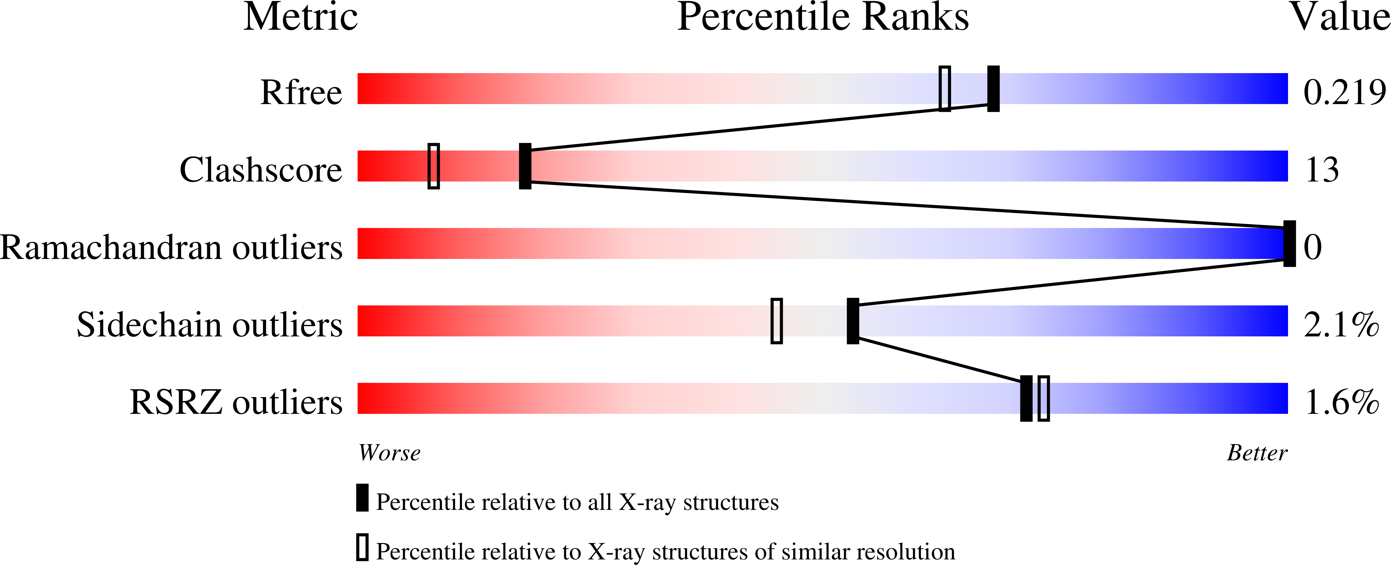
wwPDB Validation   3D Report Full Report


Ligand Structure Quality Assessment 


This is version 1.2 of the entry. See complete history


Literature

Probing the inhibitor selectivity pocket of human 20 alpha-hydroxysteroid dehydrogenase (AKR1C1) with X-ray crystallography and site-directed mutagenesis

El-Kabbani, O.Dhagat, U.Soda, M.Endo, S.Matsunaga, T.Hara, A.

(2011) Bioorg Med Chem Lett 21: 2564-2567

  • DOI: https://doi.org/10.1016/j.bmcl.2011.01.076
  • Primary Citation of Related Structures:  
    3NTY

  • PubMed Abstract: 

    Human 20α-hydroxysteroid dehydrogenase (AKR1C1) is an important drug target due to its role in the development of lung and endometrial cancers, premature birth and neuronal disorders. We report the crystal structure of AKR1C1 complexed with the first structure-based designed inhibitor 3-chloro-5-phenylsalicylic acid (K(i)=0.86 nM) bound in the active site. The binding of 3-chloro-5-phenylsalicylic acid to AKR1C1 resulted in a conformational change in the side chain of Phe311 to accommodate the bulky phenyl ring substituent at the 5-position of the inhibitor. The contributions of the nonconserved residues Leu54, Leu306, Leu308 and Phe311 to the binding were further investigated by site-directed mutagenesis, and the effects of the mutations on the K(i) value were determined. The Leu54Val and Leu306Ala mutations resulted in 6- and 81-fold increases, respectively, in K(i) values compared to the wild-type enzyme, while the remaining mutations had little or no effects.


  • Organizational Affiliation

    Medicinal Chemistry and Drug Action, Monash Institute of Pharmaceutical Sciences, Monash University, 381 Royal Parade, Parkville, Victoria 3052, Australia. [email protected]


Macromolecules
Find similar proteins by:  (by identity cutoff)  |  3D Structure
Entity ID: 1
MoleculeChains Sequence LengthOrganismDetailsImage
Aldo-keto reductase family 1 member C1323Homo sapiensMutation(s): 0 
EC: 1.1.1 (PDB Primary Data), 1.1.1.149 (PDB Primary Data), 1.3.1.20 (PDB Primary Data), 1.1.1.112 (PDB Primary Data), 1.1.1.53 (UniProt), 1.1.1.209 (UniProt), 1.1.1.51 (UniProt), 1.1.1.62 (UniProt), 1.1.1.357 (UniProt), 1.1.1.210 (UniProt)
UniProt & NIH Common Fund Data Resources
Find proteins for Q04828 (Homo sapiens)
Explore Q04828 
Go to UniProtKB:  Q04828
PHAROS:  Q04828
GTEx:  ENSG00000187134 
Entity Groups  
Sequence Clusters30% Identity50% Identity70% Identity90% Identity95% Identity100% Identity
UniProt GroupQ04828
Sequence Annotations
Expand
  • Reference Sequence
Small Molecules
Binding Affinity Annotations 
IDSourceBinding Affinity
5P3 BindingDB:  3NTY Ki: min: 0.85, max: 70 (nM) from 8 assay(s)
IC50: 100 (nM) from 1 assay(s)
Experimental Data & Validation

Experimental Data

  • Method: X-RAY DIFFRACTION
  • Resolution: 1.87 Å
  • R-Value Free: 0.253 
  • R-Value Work: 0.185 
  • R-Value Observed: 0.188 
  • Space Group: P 1 21 1
Unit Cell:
Length ( Å )Angle ( ˚ )
a = 39.607α = 90
b = 84.052β = 91.41
c = 49.051γ = 90
Software Package:
Software NamePurpose
SCALEPACKdata scaling
MOLREPphasing
REFMACrefinement
PDB_EXTRACTdata extraction
MAR345dtbdata collection
HKL-2000data reduction

Structure Validation

View Full Validation Report


Ligand Structure Quality Assessment 


Entry History 

Deposition Data

Revision History  (Full details and data files)

  • Version 1.0: 2011-04-13
    Type: Initial release
  • Version 1.1: 2011-07-13
    Changes: Version format compliance
  • Version 1.2: 2023-11-01
    Changes: Data collection, Database references, Derived calculations, Refinement description