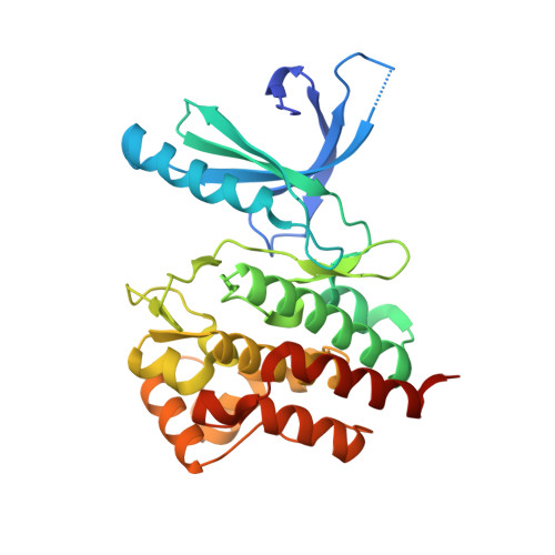
3NYX

Non-phosphorylated TYK2 JH1 domain with Quinoline-Thiadiazole-Thiophene Inhibitor


Experimental Data Snapshot

  • Method: X-RAY DIFFRACTION
  • Resolution: 2.50 Å
  • R-Value Free: 0.275 
  • R-Value Work: 0.215 
  • R-Value Observed: 0.219 

Starting Model: experimental
View more details

wwPDB Validation   3D Report Full Report


Ligand Structure Quality Assessment 


This is version 1.2 of the entry. See complete history


Literature

A new regulatory switch in a JAK protein kinase.

Tsui, V.Gibbons, P.Ultsch, M.Mortara, K.Chang, C.Blair, W.Pulk, R.Stanley, M.Starovasnik, M.Williams, D.Lamers, M.Leonard, P.Magnuson, S.Liang, J.Eigenbrot, C.

(2011) Proteins 79: 393-401

  • DOI: https://doi.org/10.1002/prot.22889
  • Primary Citation of Related Structures:  
    3NYX, 3NZ0

  • PubMed Abstract: 

    Members of the JAK family of protein kinases mediate signal transduction from cytokine receptors to transcription factor activation. Over-stimulation of these pathways is causative in immune disorders like rheumatoid arthritis, psoriasis, lupus, and Crohn's disease. A search for selective inhibitors of a JAK kinase has led to our characterization of a previously unknown kinase conformation arising from presentation of Tyr962 of TYK2 to an inhibitory small molecule via an H-bonding interaction. A small minority of protein kinase domains has a Tyrosine residue in this position within the αC-β4 loop, and it is the only amino acid commonly seen here with H-bonding potential. These discoveries will aid design of inhibitors that discriminate among the JAK family and more widely among protein kinases.


  • Organizational Affiliation

    Department of Discovery Chemistry, Genentech, Inc, South San Francisco, California 94080, USA.


Macromolecules
Find similar proteins by:  (by identity cutoff)  |  3D Structure
Entity ID: 1
MoleculeChains Sequence LengthOrganismDetailsImage
Non-receptor tyrosine-protein kinase TYK2302Homo sapiensMutation(s): 1 
Gene Names: TYK2
EC: 2.7.10.2
UniProt & NIH Common Fund Data Resources
Find proteins for P29597 (Homo sapiens)
Explore P29597 
Go to UniProtKB:  P29597
PHAROS:  P29597
GTEx:  ENSG00000105397 
Entity Groups  
Sequence Clusters30% Identity50% Identity70% Identity90% Identity95% Identity100% Identity
UniProt GroupP29597
Sequence Annotations
Expand
  • Reference Sequence
Small Molecules
Binding Affinity Annotations 
IDSourceBinding Affinity
TZ1 PDBBind:  3NYX Ki: 32 (nM) from 1 assay(s)
Experimental Data & Validation

Experimental Data

  • Method: X-RAY DIFFRACTION
  • Resolution: 2.50 Å
  • R-Value Free: 0.275 
  • R-Value Work: 0.215 
  • R-Value Observed: 0.219 
  • Space Group: P 21 21 21
Unit Cell:
Length ( Å )Angle ( ˚ )
a = 55.318α = 90
b = 70.436β = 90
c = 86.488γ = 90
Software Package:
Software NamePurpose
BOSdata collection
PHASERphasing
REFMACrefinement
HKL-2000data reduction
SCALEPACKdata scaling

Structure Validation

View Full Validation Report


Ligand Structure Quality Assessment 


Entry History 

Deposition Data

Revision History  (Full details and data files)

  • Version 1.0: 2010-10-20
    Type: Initial release
  • Version 1.1: 2011-07-13
    Changes: Version format compliance
  • Version 1.2: 2023-09-06
    Changes: Data collection, Database references, Derived calculations, Refinement description