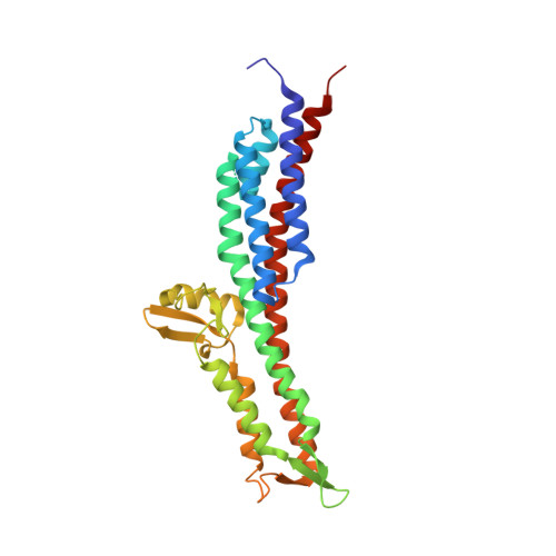
3O02

The Crystal Structure of the Salmonella Type III Secretion System Tip Protein SipD in Complex with Chenodeoxycholate


Experimental Data Snapshot

  • Method: X-RAY DIFFRACTION
  • Resolution: 1.90 Å
  • R-Value Free: 0.244 
  • R-Value Work: 0.197 
  • R-Value Observed: 0.199 

wwPDB Validation   3D Report Full Report


Ligand Structure Quality Assessment 


This is version 1.2 of the entry. See complete history


Literature

The crystal structures of the Salmonella type III secretion system tip protein SipD in complex with deoxycholate and chenodeoxycholate.

Chatterjee, S.Zhong, D.Nordhues, B.A.Battaile, K.P.Lovell, S.De Guzman, R.N.

(2011) Protein Sci 20: 75-86

  • DOI: https://doi.org/10.1002/pro.537
  • Primary Citation of Related Structures:  
    3NZZ, 3O00, 3O01, 3O02

  • PubMed Abstract: 

    The type III secretion system (T3SS) is a protein injection nanomachinery required for virulence by many human pathogenic bacteria including Salmonella and Shigella. An essential component of the T3SS is the tip protein and the Salmonella SipD and the Shigella IpaD tip proteins interact with bile salts, which serve as environmental sensors for these enteric pathogens. SipD and IpaD have long central coiled coils and their N-terminal regions form α-helical hairpins and a short helix α3 that pack against the coiled coil. Using AutoDock, others have predicted that the bile salt deoxycholate binds IpaD in a cleft formed by the α-helical hairpin and its long central coiled coil. NMR chemical shift mapping, however, indicated that the SipD residues most affected by bile salts are located in a disordered region near helix α3. Thus, how bile salts interact with SipD and IpaD is unclear. Here, we report the crystal structures of SipD in complex with the bile salts deoxycholate and chenodeoxycholate. Bile salts bind SipD in a region different from what was predicted for IpaD. In SipD, bile salts bind part of helix α3 and the C-terminus of the long central coiled coil, towards the C-terminus of the protein. We discuss the biological implication of the differences in how bile salts interact with SipD and IpaD.


  • Organizational Affiliation

    Department of Molecular Biosciences, University of Kansas, Lawrence, Kansas 66045, USA.


Macromolecules
Find similar proteins by:  (by identity cutoff)  |  3D Structure
Entity ID: 1
MoleculeChains Sequence LengthOrganismDetailsImage
Cell invasion protein sipD
A, B
308Salmonella enterica subsp. enterica serovar TyphimuriumMutation(s): 0 
Gene Names: sipDsspDSTM2883
UniProt
Find proteins for Q56026 (Salmonella typhimurium (strain LT2 / SGSC1412 / ATCC 700720))
Explore Q56026 
Go to UniProtKB:  Q56026
Entity Groups  
Sequence Clusters30% Identity50% Identity70% Identity90% Identity95% Identity100% Identity
UniProt GroupQ56026
Sequence Annotations
Expand
  • Reference Sequence
Experimental Data & Validation

Experimental Data

  • Method: X-RAY DIFFRACTION
  • Resolution: 1.90 Å
  • R-Value Free: 0.244 
  • R-Value Work: 0.197 
  • R-Value Observed: 0.199 
  • Space Group: C 1 2 1
Unit Cell:
Length ( Å )Angle ( ˚ )
a = 202.216α = 90
b = 52.37β = 90.2
c = 57.316γ = 90
Software Package:
Software NamePurpose
HKL-2000data collection
MOLREPphasing
REFMACrefinement
HKL-2000data reduction
HKL-2000data scaling

Structure Validation

View Full Validation Report


Ligand Structure Quality Assessment 


Entry History 

Deposition Data

Revision History  (Full details and data files)

  • Version 1.0: 2010-11-17
    Type: Initial release
  • Version 1.1: 2011-07-13
    Changes: Version format compliance
  • Version 1.2: 2024-02-21
    Changes: Data collection, Database references, Derived calculations