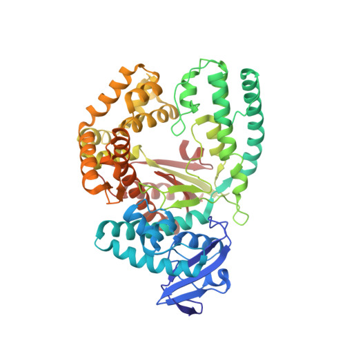
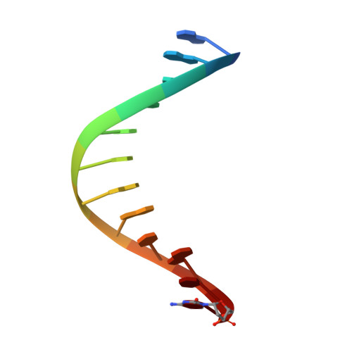
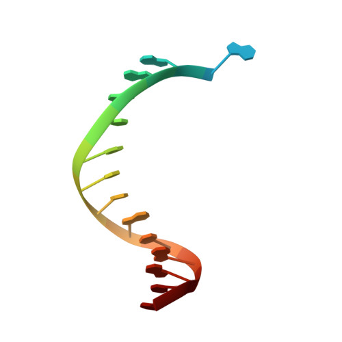
3PY8

Crystal structure of a mutant of the large fragment of DNA polymerase I from thermus aquaticus in a closed ternary complex with DNA and ddCTP


Experimental Data Snapshot

  • Method: X-RAY DIFFRACTION
  • Resolution: 1.74 Å
  • R-Value Free: 0.199 
  • R-Value Work: 0.169 
  • R-Value Observed: 0.171 

wwPDB Validation   3D Report Full Report


Ligand Structure Quality Assessment 


This is version 1.2 of the entry. See complete history


Literature

Learning from Directed Evolution: Thermus aquaticus DNA Polymerase Mutants with Translesion Synthesis Activity.

Obeid, S.Schnur, A.Gloeckner, C.Blatter, N.Welte, W.Diederichs, K.Marx, A.

(2011) Chembiochem 12: 1574-1580

  • DOI: https://doi.org/10.1002/cbic.201000783
  • Primary Citation of Related Structures:  
    3PO4, 3PO5, 3PY8

  • PubMed Abstract: 

    DNA is being constantly damaged by endo- and exogenous agents such as reactive oxygen species, chemicals, radioactivity, and ultraviolet radiation. Additionally, DNA is inherently labile, and this can result in, for example, the spontaneous hydrolysis of the glycosidic bond that connects the sugar and the nucleobase moieties in DNA; this results in abasic sites. It has long been obscure how cells achieve DNA synthesis past these lesions, and only recently has it been discovered that several specialized DNA polymerases are involved in translesion synthesis. The underlying mechanisms that render one DNA polymerase competent in translesion synthesis while another DNA polymerase fails are still indistinct. Recently two variants of Taq DNA polymerase that exhibited higher lesion bypass ability than the wild-type enzyme were identified by directed-evolution approaches. Strikingly, in both approaches it was independently found that substitution of a single nonpolar amino acid side chain by a cationic side chain increases the capability of translesion synthesis. Here, we combined both mutations in a single enzyme. We found that the KlenTaq DNA polymerase that bore both mutations superseded the wild-type as well as the respective single mutants in translesion-bypass proficiency. Further insights in the molecular basis of the detected gain of translesion-synthesis function were obtained by structural studies of DNA polymerase variants caught in processing canonical and damaged substrates. We found that increased positive charge of the surface potential in the area proximal to the negatively charged substrates promotes translesion synthesis by KlenTaq DNA polymerase, an enzyme that has very limited naturally evolved capability to perform translesion synthesis. Since expanded positively charged surface potential areas are also found in naturally evolved translesion DNA polymerases, our results underscore the impact of charge on the proficiency of naturally evolved translesion DNA polymerases.


  • Organizational Affiliation

    Department of Chemistry, Konstanz Research School Chemical Biology, University of Konstanz, Konstanz, Germany.


Macromolecules

Find similar proteins by:  (by identity cutoff)  |  3D Structure
Entity ID: 1
MoleculeChains Sequence LengthOrganismDetailsImage
DNA polymerase I540Thermus aquaticusMutation(s): 2 
Gene Names: polApol1
EC: 2.7.7.7
UniProt
Find proteins for P19821 (Thermus aquaticus)
Explore P19821 
Go to UniProtKB:  P19821
Entity Groups  
Sequence Clusters30% Identity50% Identity70% Identity90% Identity95% Identity100% Identity
UniProt GroupP19821
Sequence Annotations
Expand
  • Reference Sequence

Find similar nucleic acids by:  Sequence   |   3D Structure  

Entity ID: 2
MoleculeChains LengthOrganismImage
DNA (5'-D(*GP*AP*CP*CP*AP*CP*GP*GP*CP*GP*CP*(DOC))-3')12N/A
Sequence Annotations
Expand
  • Reference Sequence

Find similar nucleic acids by:  Sequence   |   3D Structure  

Entity ID: 3
MoleculeChains LengthOrganismImage
DNA (5'-D(*AP*A*AP*GP*GP*GP*CP*GP*CP*CP*GP*TP*GP*GP*TP*C)-3')16N/A
Sequence Annotations
Expand
  • Reference Sequence
Small Molecules
Ligands 4 Unique
IDChains Name / Formula / InChI Key2D Diagram3D Interactions
DCT
Query on DCT

Download Ideal Coordinates CCD File 
D [auth A]2',3'-DIDEOXYCYTIDINE 5'-TRIPHOSPHATE
C9 H16 N3 O12 P3
ARLKCWCREKRROD-POYBYMJQSA-N
GOL
Query on GOL

Download Ideal Coordinates CCD File 
E [auth A],
J [auth A]
GLYCEROL
C3 H8 O3
PEDCQBHIVMGVHV-UHFFFAOYSA-N
MN
Query on MN

Download Ideal Coordinates CCD File 
F [auth A],
H [auth A],
I [auth A],
K [auth B],
L [auth C]
MANGANESE (II) ION
Mn
WAEMQWOKJMHJLA-UHFFFAOYSA-N
MG
Query on MG

Download Ideal Coordinates CCD File 
G [auth A]MAGNESIUM ION
Mg
JLVVSXFLKOJNIY-UHFFFAOYSA-N
Experimental Data & Validation

Experimental Data

  • Method: X-RAY DIFFRACTION
  • Resolution: 1.74 Å
  • R-Value Free: 0.199 
  • R-Value Work: 0.169 
  • R-Value Observed: 0.171 
  • Space Group: P 31 2 1
Unit Cell:
Length ( Å )Angle ( ˚ )
a = 108.61α = 90
b = 108.61β = 90
c = 90.57γ = 120
Software Package:
Software NamePurpose
ADSCdata collection
PHENIXmodel building
PHENIXrefinement
XDSdata reduction
XDSdata scaling
PHENIXphasing

Structure Validation

View Full Validation Report


Ligand Structure Quality Assessment 


Entry History 

Deposition Data

Revision History  (Full details and data files)

  • Version 1.0: 2011-06-15
    Type: Initial release
  • Version 1.1: 2011-07-13
    Changes: Version format compliance
  • Version 1.2: 2024-02-21
    Changes: Data collection, Database references, Derived calculations