3QWH

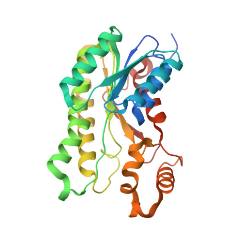
Crystal structure of the 17beta-hydroxysteroid dehydrogenase from Cochliobolus lunatus in complex with NADPH and kaempferol


Experimental Data Snapshot

  • Method: X-RAY DIFFRACTION
  • Resolution: 2.62 Å
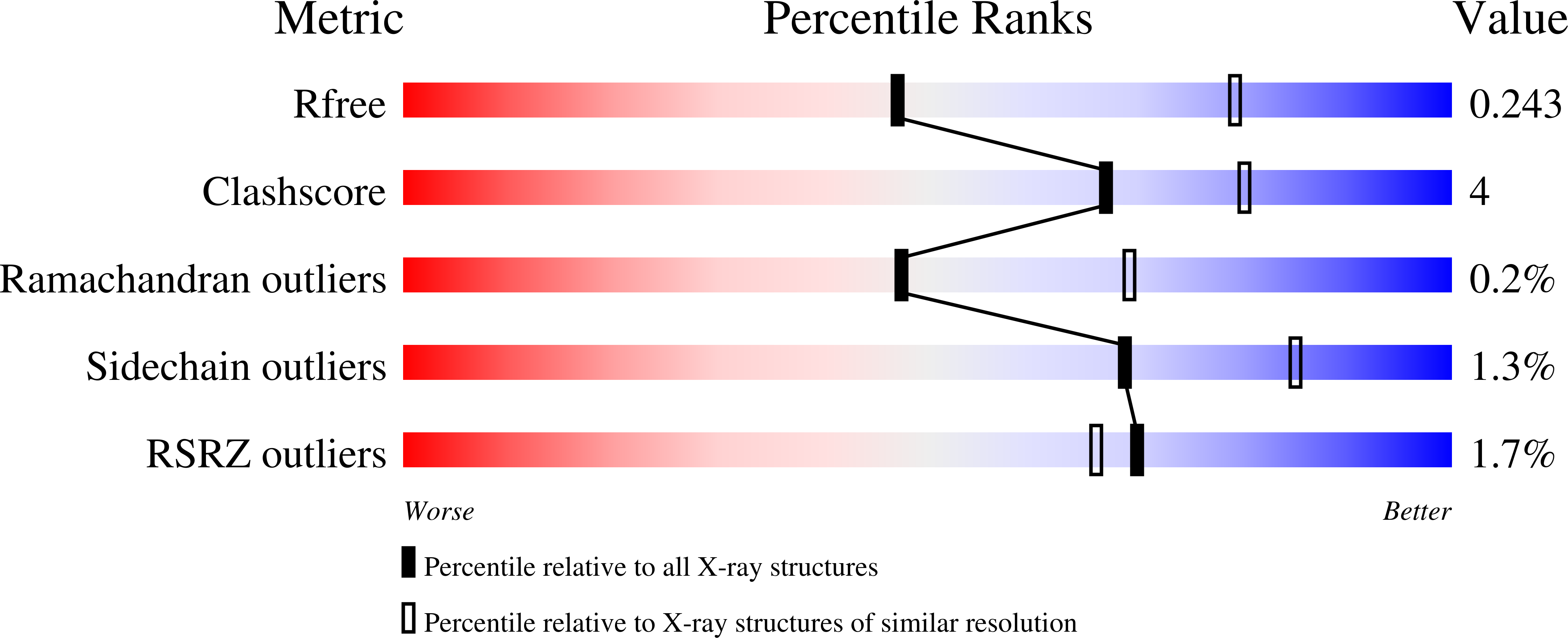
  • R-Value Free: 0.240 
  • R-Value Work: 0.168 
  • R-Value Observed: 0.172 

Starting Model: experimental
View more details

wwPDB Validation   3D Report Full Report


Ligand Structure Quality Assessment 


This is version 1.3 of the entry. See complete history


Literature

Structural basis for inhibition of 17 beta-hydroxysteroid dehydrogenases by phytoestrogens: The case of fungal 17 beta-HSDcl.

Cassetta, A.Stojan, J.Krastanova, I.Kristan, K.Brunskole Svegelj, M.Lamba, D.Rizner, T.L.

(2017) J Steroid Biochem Mol Biol 171: 80-93

  • DOI: https://doi.org/10.1016/j.jsbmb.2017.02.020
  • Primary Citation of Related Structures:  
    3QWH, 4FJ0, 4FJ1, 4FJ2

  • PubMed Abstract: 

    Phytoestrogens are plant-derived compounds that functionally and structurally mimic mammalian estrogens. Phytoestrogens have broad inhibitory activities toward several steroidogenic enzymes, such as the 17β-hydroxysteroid dehydrogenases (17β-HSDs), which modulate the biological potency of androgens and estrogens in mammals. However, to date, no crystallographic data are available to explain phytoestrogens binding to mammalian 17β-HSDs. NADP(H)-dependent 17β-HSD from the filamentous fungus Cochliobolus lunatus (17β-HSDcl) has been the subject of extensive biochemical, kinetic and quantitative structure-activity relationship studies that have shown that the flavonols are the most potent inhibitors. In the present study, we investigated the structure-activity relationships of the ternary complexes between the holo form of 17β-HSDcl and the flavonols kaempferol and 3,7-dihydroxyflavone, in comparison with the isoflavones genistein and biochanin A. Crystallographic data are accompanied by kinetic analysis of the inhibition mechanisms for six flavonols (3-hydroxyflavone, 3,7-dihydroxyflavone, kaempferol, quercetin, fisetin, myricetin), one flavanone (naringenin), one flavone (luteolin), and two isoflavones (genistein, biochanin A). The kinetics analysis shows that the degree of hydroxylation of ring B significantly influences the overall inhibitory efficacy of the flavonols. A distinct binding mode defines the interactions between 17β-HSDcl and the flavones and isoflavones. Moreover, the complex with biochanin A reveals an unusual binding mode that appears to account for its greater inhibition of 17β-HSDcl with respect to genistein. Overall, these data provide a blueprint for identification of the distinct molecular determinants that underpin 17β-HSD inhibition by phytoestrogens.


  • Organizational Affiliation

    Istituto di Cristallografia, UOS Trieste, Consiglio Nazionale delle Ricerche, S. S. 14-Km 163.5, I-34149, Trieste, Italy. Electronic address: [email protected].


Macromolecules
Find similar proteins by:  (by identity cutoff)  |  3D Structure
Entity ID: 1
MoleculeChains Sequence LengthOrganismDetailsImage
17beta-hydroxysteroid dehydrogenase
A, B, C, D
270Curvularia lunataMutation(s): 0 
Gene Names: 17HSDcl
EC: 1.1.1.62
UniProt
Find proteins for O93874 (Cochliobolus lunatus)
Explore O93874 
Go to UniProtKB:  O93874
Entity Groups  
Sequence Clusters30% Identity50% Identity70% Identity90% Identity95% Identity100% Identity
UniProt GroupO93874
Sequence Annotations
Expand
  • Reference Sequence
Small Molecules
Ligands 3 Unique
IDChains Name / Formula / InChI Key2D Diagram3D Interactions
NAP
Query on NAP

Download Ideal Coordinates CCD File 
E [auth A],
G [auth B],
J [auth C],
K [auth D]
NADP NICOTINAMIDE-ADENINE-DINUCLEOTIDE PHOSPHATE
C21 H28 N7 O17 P3
XJLXINKUBYWONI-NNYOXOHSSA-N
KMP
Query on KMP

Download Ideal Coordinates CCD File 
F [auth A],
H [auth B],
L [auth D]
3,5,7-TRIHYDROXY-2-(4-HYDROXYPHENYL)-4H-CHROMEN-4-ONE
C15 H10 O6
IYRMWMYZSQPJKC-UHFFFAOYSA-N
PEG
Query on PEG

Download Ideal Coordinates CCD File 
I [auth B]DI(HYDROXYETHYL)ETHER
C4 H10 O3
MTHSVFCYNBDYFN-UHFFFAOYSA-N
Experimental Data & Validation

Experimental Data

  • Method: X-RAY DIFFRACTION
  • Resolution: 2.62 Å
  • R-Value Free: 0.240 
  • R-Value Work: 0.168 
  • R-Value Observed: 0.172 
  • Space Group: P 1 21 1
Unit Cell:
Length ( Å )Angle ( ˚ )
a = 62.95α = 90
b = 116.64β = 102.54
c = 70.24γ = 90
Software Package:
Software NamePurpose
MAR345data collection
MOLREPphasing
REFMACrefinement
MOSFLMdata reduction
SCALAdata scaling

Structure Validation

View Full Validation Report


Ligand Structure Quality Assessment 


Entry History 

Deposition Data

Revision History  (Full details and data files)

  • Version 1.0: 2012-03-21
    Type: Initial release
  • Version 1.1: 2017-07-19
    Changes: Database references
  • Version 1.2: 2017-11-08
    Changes: Refinement description
  • Version 1.3: 2023-09-13
    Changes: Data collection, Database references, Derived calculations, Refinement description