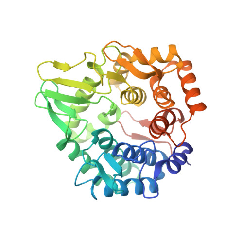
3QXQ

Structure of the bacterial cellulose synthase subunit Z in complex with cellopentaose


Experimental Data Snapshot

  • Method: X-RAY DIFFRACTION
  • Resolution: 2.20 Å
  • R-Value Free: 0.227 
  • R-Value Work: 0.168 
  • R-Value Observed: 0.171 

wwPDB Validation   3D Report Full Report


This is version 2.1 of the entry. See complete history


Literature

Apo- and cellopentaose-bound structures of the bacterial cellulose synthase subunit BcsZ.

Mazur, O.Zimmer, J.

(2011) J Biol Chem 286: 17601-17606

  • DOI: https://doi.org/10.1074/jbc.M111.227660
  • Primary Citation of Related Structures:  
    3QXF, 3QXQ

  • PubMed Abstract: 

    Cellulose, a very abundant extracellular polysaccharide, is synthesized in a finely tuned process that involves the activity of glycosyl-transferases and hydrolases. The cellulose microfibril consists of bundles of linear β-1,4-glucan chains that are synthesized inside the cell; however, the mechanism by which these polymers traverse the cell membrane is currently unknown. In Gram-negative bacteria, the cellulose synthase complex forms a trans-envelope complex consisting of at least four subunits. Although three of these subunits account for the synthesis and translocation of the polysaccharide, the fourth subunit, BcsZ, is a periplasmic protein with endo-β-1,4-glucanase activity. BcsZ belongs to family eight of glycosyl-hydrolases, and its activity is required for optimal synthesis and membrane translocation of cellulose. In this study we report two crystal structures of BcsZ from Escherichia coli. One structure shows the wild-type enzyme in its apo form, and the second structure is for a catalytically inactive mutant of BcsZ in complex with the substrate cellopentaose. The structures demonstrate that BcsZ adopts an (α/α)(6)-barrel fold and that it binds four glucan moieties of cellopentaose via highly conserved residues exclusively on the nonreducing side of its catalytic center. Thus, the BcsZ-cellopentaose structure most likely represents a posthydrolysis state in which the newly formed nonreducing end has already left the substrate binding pocket while the enzyme remains attached to the truncated polysaccharide chain. We further show that BcsZ efficiently degrades β-1,4-glucans in in vitro cellulase assays with carboxymethyl-cellulose as substrate.


  • Organizational Affiliation

    Center for Membrane Biology, Department of Molecular Physiology and Biological Physics, University of Virginia, Charlottesville, Virginia 22908, USA.


Macromolecules
Find similar proteins by:  (by identity cutoff)  |  3D Structure
Entity ID: 1
MoleculeChains Sequence LengthOrganismDetailsImage
Endoglucanase
A, B, C, D
355Escherichia coli K-12Mutation(s): 1 
Gene Names: bcsZbcsCyhjMb3531JW3499
EC: 3.2.1.4
UniProt
Find proteins for P37651 (Escherichia coli (strain K12))
Explore P37651 
Go to UniProtKB:  P37651
Entity Groups  
Sequence Clusters30% Identity50% Identity70% Identity90% Identity95% Identity100% Identity
UniProt GroupP37651
Sequence Annotations
Expand
  • Reference Sequence
Oligosaccharides

Help

Entity ID: 2
MoleculeChains Length2D Diagram Glycosylation3D Interactions
beta-D-glucopyranose-(1-4)-beta-D-glucopyranose-(1-4)-beta-D-glucopyranose-(1-4)-beta-D-glucopyranose-(1-4)-beta-D-glucopyranose
E, F, G, H
5N/A
Glycosylation Resources
GlyTouCan:  G91683DU
GlyCosmos:  G91683DU
Experimental Data & Validation

Experimental Data

  • Method: X-RAY DIFFRACTION
  • Resolution: 2.20 Å
  • R-Value Free: 0.227 
  • R-Value Work: 0.168 
  • R-Value Observed: 0.171 
  • Space Group: P 1 21 1
Unit Cell:
Length ( Å )Angle ( ˚ )
a = 90.678α = 90
b = 99.605β = 102.98
c = 93.073γ = 90
Software Package:
Software NamePurpose
HKL-2000data collection
PHASERphasing
REFMACrefinement
HKL-2000data reduction
HKL-2000data scaling

Structure Validation

View Full Validation Report


Entry History 

Deposition Data

  • Released Date: 2011-03-30 
  • Deposition Author(s): Zimmer, J.

Revision History  (Full details and data files)

  • Version 1.0: 2011-03-30
    Type: Initial release
  • Version 1.1: 2011-07-13
    Changes: Version format compliance
  • Version 1.2: 2011-08-10
    Changes: Database references
  • Version 2.0: 2020-07-29
    Type: Remediation
    Reason: Carbohydrate remediation
    Changes: Atomic model, Data collection, Database references, Derived calculations, Non-polymer description, Structure summary
  • Version 2.1: 2024-02-21
    Changes: Data collection, Database references, Structure summary