3RAE

Quinolone(Levofloxacin)-DNA cleavage complex of type IV topoisomerase from S. pneumoniae


Experimental Data Snapshot

  • Method: X-RAY DIFFRACTION
  • Resolution: 2.90 Å
  • R-Value Free: 0.226 
  • R-Value Work: 0.186 
  • R-Value Observed: 0.190 

Starting Model: experimental
View more details

wwPDB Validation   3D Report Full Report


Ligand Structure Quality Assessment 


This is version 1.3 of the entry. See complete history


Literature

Structure of a quinolone-stabilized cleavage complex of topoisomerase IV from Klebsiella pneumoniae and comparison with a related Streptococcus pneumoniae complex.

Veselkov, D.A.Laponogov, I.Pan, X.S.Selvarajah, J.Skamrova, G.B.Branstrom, A.Narasimhan, J.Prasad, J.V.Fisher, L.M.Sanderson, M.R.

(2016) Acta Crystallogr D Biol Crystallogr 72: 488-496

  • DOI: https://doi.org/10.1107/S2059798316001212
  • Primary Citation of Related Structures:  
    3RAE, 5EIX

  • PubMed Abstract: 

    Klebsiella pneumoniae is a Gram-negative bacterium that is responsible for a range of common infections, including pulmonary pneumonia, bloodstream infections and meningitis. Certain strains of Klebsiella have become highly resistant to antibiotics. Despite the vast amount of research carried out on this class of bacteria, the molecular structure of its topoisomerase IV, a type II topoisomerase essential for catalysing chromosomal segregation, had remained unknown. In this paper, the structure of its DNA-cleavage complex is reported at 3.35 Å resolution. The complex is comprised of ParC breakage-reunion and ParE TOPRIM domains of K. pneumoniae topoisomerase IV with DNA stabilized by levofloxacin, a broad-spectrum fluoroquinolone antimicrobial agent. This complex is compared with a similar complex from Streptococcus pneumoniae, which has recently been solved.


  • Organizational Affiliation

    Randall Division of Cell and Molecular Biophysics, King's College London, 3rd Floor, New Hunt's House, Guy's Campus, London SE1 1UL, England.


Macromolecules

Find similar proteins by:  (by identity cutoff)  |  3D Structure
Entity ID: 1
MoleculeChains Sequence LengthOrganismDetailsImage
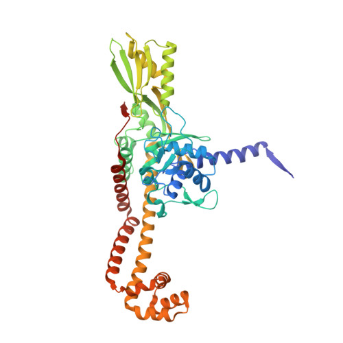
DNA topoisomerase 4 subunit A
A, B
496Streptococcus pneumoniaeMutation(s): 0 
Gene Names: parCSP_0855
EC: 5.99.1 (PDB Primary Data), 5.6.2.2 (UniProt)
UniProt
Find proteins for P72525 (Streptococcus pneumoniae serotype 4 (strain ATCC BAA-334 / TIGR4))
Explore P72525 
Go to UniProtKB:  P72525
Entity Groups  
Sequence Clusters30% Identity50% Identity70% Identity90% Identity95% Identity100% Identity
UniProt GroupP72525
Sequence Annotations
Expand
  • Reference Sequence
Find similar proteins by:  (by identity cutoff)  |  3D Structure
Entity ID: 2
MoleculeChains Sequence LengthOrganismDetailsImage
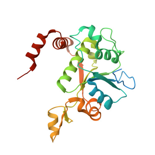
DNA topoisomerase 4 subunit B
C, D
268Streptococcus pneumoniaeMutation(s): 0 
Gene Names: parESP_0852
EC: 5.99.1 (PDB Primary Data), 5.6.2.2 (UniProt)
UniProt
Find proteins for Q59961 (Streptococcus pneumoniae serotype 4 (strain ATCC BAA-334 / TIGR4))
Explore Q59961 
Go to UniProtKB:  Q59961
Entity Groups  
Sequence Clusters30% Identity50% Identity70% Identity90% Identity95% Identity100% Identity
UniProt GroupQ59961
Sequence Annotations
Expand
  • Reference Sequence

Find similar nucleic acids by:  Sequence   |   3D Structure  

Entity ID: 3
MoleculeChains LengthOrganismImage
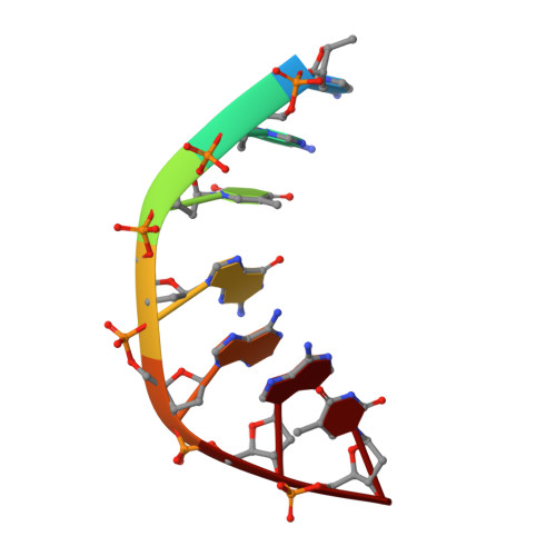
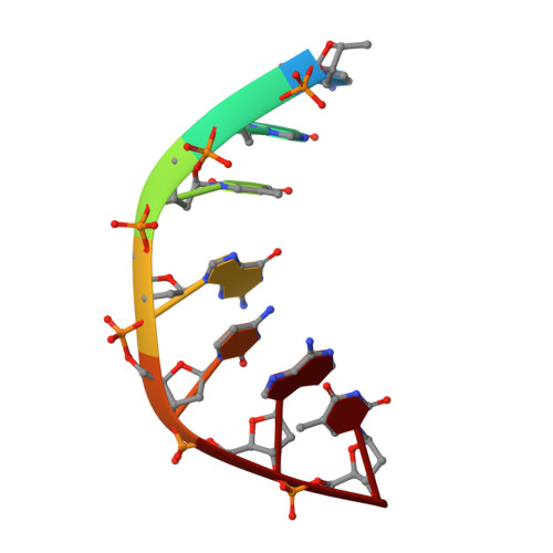
5'-D(*CP*AP*TP*GP*AP*AP*T)-3'7N/A
Sequence Annotations
Expand
  • Reference Sequence

Find similar nucleic acids by:  Sequence   |   3D Structure  

Entity ID: 4
MoleculeChains LengthOrganismImage
5'-D(P*AP*GP*TP*CP*AP*TP*TP*CP*AP*TP*G)-3'11N/A
Sequence Annotations
Expand
  • Reference Sequence

Find similar nucleic acids by:  Sequence   |   3D Structure  

Entity ID: 5
MoleculeChains LengthOrganismImage
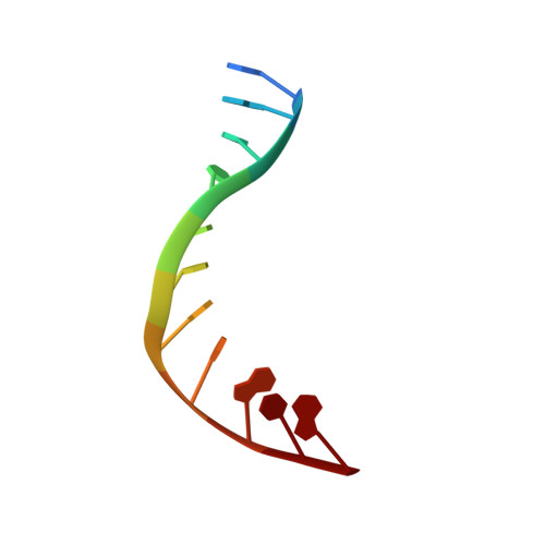
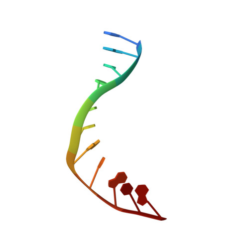
5'-D(*CP*GP*TP*GP*CP*AP*T)-3'7N/A
Sequence Annotations
Expand
  • Reference Sequence

Find similar nucleic acids by:  Sequence   |   3D Structure  

Entity ID: 6
MoleculeChains LengthOrganismImage
5'-D(P*GP*AP*CP*TP*AP*TP*GP*CP*AP*CP*G)-3'11N/A
Sequence Annotations
Expand
  • Reference Sequence
Small Molecules
Ligands 2 Unique
IDChains Name / Formula / InChI Key2D Diagram3D Interactions
LFX
Query on LFX

Download Ideal Coordinates CCD File 
N [auth F],
P [auth H]
(3S)-9-fluoro-3-methyl-10-(4-methylpiperazin-1-yl)-7-oxo-2,3-dihydro-7H-[1,4]oxazino[2,3,4-ij]quinoline-6-carboxylic acid
C18 H20 F N3 O4
GSDSWSVVBLHKDQ-JTQLQIEISA-N
MG
Query on MG

Download Ideal Coordinates CCD File 
I [auth A]
J [auth A]
K [auth B]
L [auth C]
M [auth D]
I [auth A],
J [auth A],
K [auth B],
L [auth C],
M [auth D],
O [auth G]
MAGNESIUM ION
Mg
JLVVSXFLKOJNIY-UHFFFAOYSA-N
Experimental Data & Validation

Experimental Data

  • Method: X-RAY DIFFRACTION
  • Resolution: 2.90 Å
  • R-Value Free: 0.226 
  • R-Value Work: 0.186 
  • R-Value Observed: 0.190 
  • Space Group: P 31 2 1
Unit Cell:
Length ( Å )Angle ( ˚ )
a = 157.831α = 90
b = 157.831β = 90
c = 211.147γ = 120
Software Package:
Software NamePurpose
GDEdata collection
PHASERphasing
PHENIXrefinement
HKL-2000data reduction
HKL-2000data scaling

Structure Validation

View Full Validation Report


Ligand Structure Quality Assessment 


Entry History 

Deposition Data

Revision History  (Full details and data files)

  • Version 1.0: 2012-04-25
    Type: Initial release
  • Version 1.1: 2016-04-20
    Changes: Database references
  • Version 1.2: 2023-11-01
    Changes: Data collection, Database references, Derived calculations, Refinement description
  • Version 1.3: 2024-10-30
    Changes: Structure summary