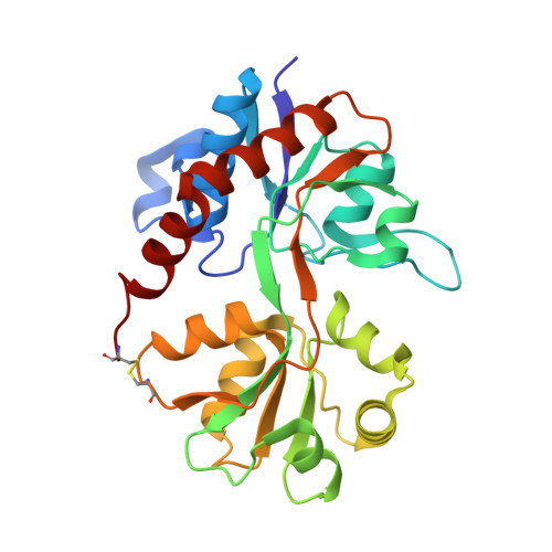
3RTW

Nitrowillardiine bound to the ligand binding domain of GluA2


Experimental Data Snapshot

  • Method: X-RAY DIFFRACTION
  • Resolution: 2.10 Å
  • R-Value Free: 0.259 
  • R-Value Work: 0.205 
  • R-Value Observed: 0.207 

Starting Model: experimental
View more details

wwPDB Validation   3D Report Full Report


Ligand Structure Quality Assessment 


This is version 1.4 of the entry. See complete history


Literature

Mechanisms of Modal Activation of GluA3 Receptors.

Poon, K.Ahmed, A.H.Nowak, L.M.Oswald, R.E.

(2011) Mol Pharmacol 80: 49-59

  • DOI: https://doi.org/10.1124/mol.111.071688
  • Primary Citation of Related Structures:  
    3RT6, 3RT8, 3RTF, 3RTW

  • PubMed Abstract: 

    AMPA receptors are the major excitatory neurotransmitter receptors in the central nervous system and are involved in numerous neurological disorders. An agonist-binding site is present in each of four subunits that form a functional channel. Binding consists of three steps: docking of agonist to the bilobed ligand binding domain (LBD), closure of the LBD, and increased stability of the closed-lobe conformation through interlobe hydrogen bonding. We describe GluA3 single channel currents activated by nitrowillardiine (NO(2)W) and chlorowillardiine (ClW) in the presence of cyclothiazide, in conjunction with crystal structures of GluA2 and GluA3 LBDs bound to fluorowillardiine (FW), ClW, and NO(2)W. When bound to NO(2)W or ClW, the GluA3 channel opens to three conductance levels with comparable open probabilities and displays modal behavior similar to that obtained with glutamate and FW as agonists (Poon et al., 2010). At lower concentrations, ClW evoked an alternate kinetic behavior, consisting of high open probability in lower conductance states. The structure of ClW bound to GluA3 LBD exhibits a unique partially open hydrogen bonding structure that may be associated with these alternative kinetics. NO(2)W evoked longer open times than seen for other agonists in high and very high modes. The structure ofGluA2 LBD bound to NO(2)W exhibits fully closed lobes with additional interlobe interactions mediated by the nitro group. Beyond differences in efficacy between full and partial agonists, the complexities of the single channel behavior of AMPA receptors may also be associated with small interactions that modify the stability of various degrees of closure.


  • Organizational Affiliation

    Department of Molecular Medicine, Cornell University, Ithaca, NY 14853, USA.


Macromolecules
Find similar proteins by:  (by identity cutoff)  |  3D Structure
Entity ID: 1
MoleculeChains Sequence LengthOrganismDetailsImage
Glutamate receptor 2A [auth B],
B [auth D],
C [auth F]
258Rattus norvegicusMutation(s): 0 
Gene Names: Glur2Gria2
UniProt
Find proteins for P19491 (Rattus norvegicus)
Explore P19491 
Go to UniProtKB:  P19491
Entity Groups  
Sequence Clusters30% Identity50% Identity70% Identity90% Identity95% Identity100% Identity
UniProt GroupP19491
Sequence Annotations
Expand
  • Reference Sequence
Small Molecules
Ligands 2 Unique
IDChains Name / Formula / InChI Key2D Diagram3D Interactions
NWD
Query on NWD

Download Ideal Coordinates CCD File 
D [auth B],
G [auth D],
I [auth F]
3-(5-nitro-2,4-dioxo-3,4-dihydropyrimidin-1(2H)-yl)-L-alanine
C7 H8 N4 O6
IEBVITXSHAFLJR-VKHMYHEASA-N
ZN
Query on ZN

Download Ideal Coordinates CCD File 
E [auth B],
F [auth B],
H [auth D],
J [auth F],
K [auth F]
ZINC ION
Zn
PTFCDOFLOPIGGS-UHFFFAOYSA-N
Experimental Data & Validation

Experimental Data

  • Method: X-RAY DIFFRACTION
  • Resolution: 2.10 Å
  • R-Value Free: 0.259 
  • R-Value Work: 0.205 
  • R-Value Observed: 0.207 
  • Space Group: P 2 21 21
Unit Cell:
Length ( Å )Angle ( ˚ )
a = 48.077α = 90
b = 114.102β = 90
c = 165.048γ = 90
Software Package:
Software NamePurpose
HKL-2000data collection
PHENIXmodel building
PHENIXrefinement
HKL-2000data reduction
HKL-2000data scaling
PHENIXphasing

Structure Validation

View Full Validation Report


Ligand Structure Quality Assessment 


Entry History 

Deposition Data

Revision History  (Full details and data files)

  • Version 1.0: 2011-05-18
    Type: Initial release
  • Version 1.1: 2011-07-13
    Changes: Version format compliance
  • Version 1.2: 2017-07-26
    Changes: Data collection, Source and taxonomy
  • Version 1.3: 2023-09-13
    Changes: Data collection, Database references, Derived calculations, Refinement description
  • Version 1.4: 2024-10-16
    Changes: Structure summary