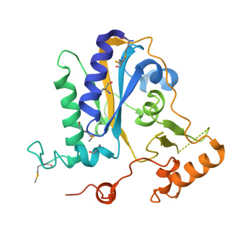
3SBY

Crystal Structure of SeMet-Substituted Apo-MMACHC (1-244), a human B12 processing enzyme


Experimental Data Snapshot

  • Method: X-RAY DIFFRACTION
  • Resolution: 2.71 Å
  • R-Value Free: 0.290 
  • R-Value Work: 0.228 
  • R-Value Observed: 0.231 

wwPDB Validation   3D Report Full Report


This is version 1.5 of the entry. See complete history


Literature

Structural basis of multifunctionality in a vitamin B12-processing enzyme.

Koutmos, M.Gherasim, C.Smith, J.L.Banerjee, R.

(2011) J Biol Chem 286: 29780-29787

  • DOI: https://doi.org/10.1074/jbc.M111.261370
  • Primary Citation of Related Structures:  
    3SBY, 3SBZ, 3SC0

  • PubMed Abstract: 

    An early step in the intracellular processing of vitamin B(12) involves CblC, which exhibits dual reactivity, catalyzing the reductive decyanation of cyanocobalamin (vitamin B(12)), and the dealkylation of alkylcobalamins (e.g. methylcobalamin; MeCbl). Insights into how the CblC scaffold supports this chemical dichotomy have been unavailable despite it being the most common locus of patient mutations associated with inherited cobalamin disorders that manifest in both severe homocystinuria and methylmalonic aciduria. Herein, we report structures of human CblC, with and without bound MeCbl, which provide novel biochemical insights into its mechanism of action. Our results reveal that CblC is the most divergent member of the NADPH-dependent flavin reductase family and can use FMN or FAD as a prosthetic group to catalyze reductive decyanation. Furthermore, CblC is the first example of an enzyme with glutathione transferase activity that has a sequence and structure unrelated to the GST superfamily. CblC thus represents an example of evolutionary adaptation of a common structural platform to perform diverse chemistries. The CblC structure allows us to rationalize the biochemical basis of a number of pathological mutations associated with severe clinical phenotypes.


  • Organizational Affiliation

    Department of Biological Chemistry and the Life Sciences Institute, University of Michigan Medical Center, Ann Arbor, Michigan 48109-0600, USA.


Macromolecules
Find similar proteins by:  (by identity cutoff)  |  3D Structure
Entity ID: 1
MoleculeChains Sequence LengthOrganismDetailsImage
Methylmalonic aciduria and homocystinuria type C protein
A, B
252Homo sapiensMutation(s): 4 
Gene Names: MMACHC
EC: 2.5.1.151 (UniProt), 1.16.1.6 (UniProt)
UniProt & NIH Common Fund Data Resources
Find proteins for Q9Y4U1 (Homo sapiens)
Explore Q9Y4U1 
Go to UniProtKB:  Q9Y4U1
PHAROS:  Q9Y4U1
GTEx:  ENSG00000132763 
Entity Groups  
Sequence Clusters30% Identity50% Identity70% Identity90% Identity95% Identity100% Identity
UniProt GroupQ9Y4U1
Sequence Annotations
Expand
  • Reference Sequence
Small Molecules
Modified Residues  1 Unique
IDChains TypeFormula2D DiagramParent
MSE
Query on MSE
A, B
L-PEPTIDE LINKINGC5 H11 N O2 SeMET
Experimental Data & Validation

Experimental Data

  • Method: X-RAY DIFFRACTION
  • Resolution: 2.71 Å
  • R-Value Free: 0.290 
  • R-Value Work: 0.228 
  • R-Value Observed: 0.231 
  • Space Group: P 65
Unit Cell:
Length ( Å )Angle ( ˚ )
a = 69.267α = 90
b = 69.267β = 90
c = 201.848γ = 120
Software Package:
Software NamePurpose
DENZOdata reduction
SCALEPACKdata scaling
SOLVEphasing
RESOLVEphasing
REFMACrefinement
PDB_EXTRACTdata extraction
Blu-Icedata collection
HKL-2000data reduction
HKL-2000data scaling

Structure Validation

View Full Validation Report


Entry History 

Deposition Data

Revision History  (Full details and data files)

  • Version 1.0: 2011-06-22
    Type: Initial release
  • Version 1.1: 2011-07-13
    Changes: Version format compliance
  • Version 1.2: 2011-08-10
    Changes: Database references
  • Version 1.3: 2012-04-25
    Changes: Database references
  • Version 1.4: 2017-11-08
    Changes: Refinement description
  • Version 1.5: 2024-11-06
    Changes: Data collection, Database references, Derived calculations, Structure summary