3SE2

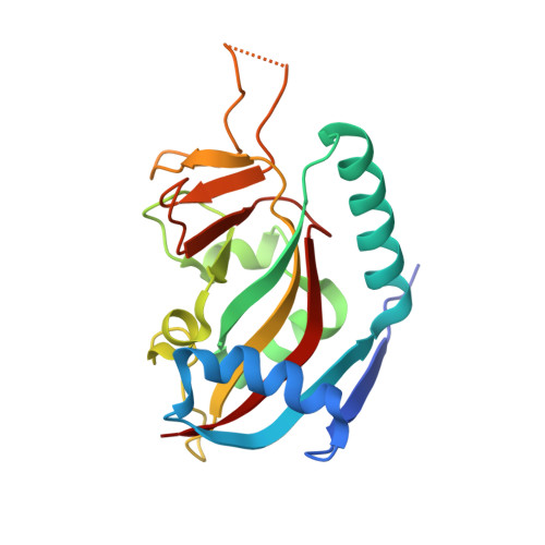
Human poly(ADP-ribose) polymerase 14 (PARP14/ARTD8) - catalytic domain in complex with 6(5H)-phenanthridinone


Experimental Data Snapshot

  • Method: X-RAY DIFFRACTION
  • Resolution: 2.30 Å
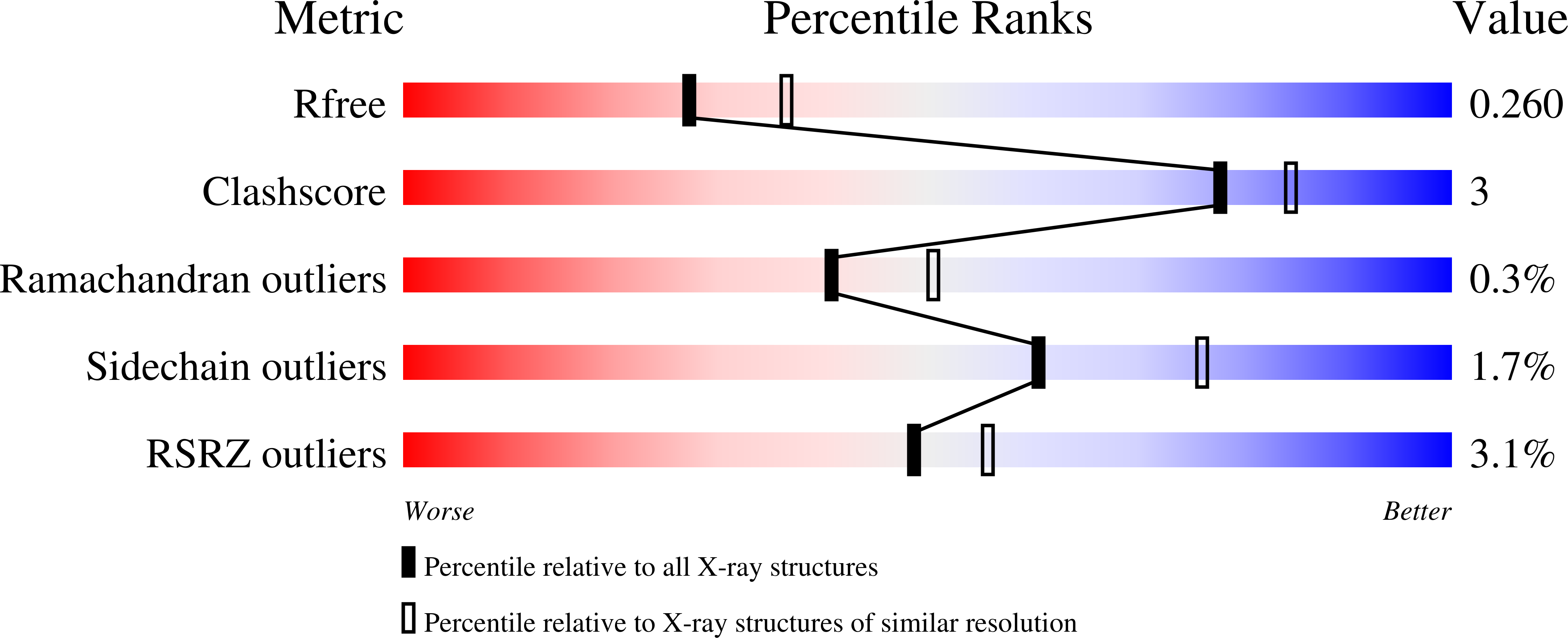
  • R-Value Free: 0.255 
  • R-Value Work: 0.208 
  • R-Value Observed: 0.210 

Starting Model: experimental
View more details

wwPDB Validation   3D Report Full Report


Ligand Structure Quality Assessment 


This is version 1.6 of the entry. See complete history


Literature

Family-wide chemical profiling and structural analysis of PARP and tankyrase inhibitors.

Wahlberg, E.Karlberg, T.Kouznetsova, E.Markova, N.Macchiarulo, A.Thorsell, A.G.Pol, E.Frostell, A.Ekblad, T.Oncu, D.Kull, B.Robertson, G.M.Pellicciari, R.Schuler, H.Weigelt, J.

(2012) Nat Biotechnol 30: 283-288

  • DOI: https://doi.org/10.1038/nbt.2121
  • Primary Citation of Related Structures:  
    3GOY, 3MHJ, 3MHK, 3P0N, 3P0P, 3P0Q, 3SE2, 3SMI, 3SMJ

  • PubMed Abstract: 

    Inhibitors of poly-ADP-ribose polymerase (PARP) family proteins are currently in clinical trials as cancer therapeutics, yet the specificity of many of these compounds is unknown. Here we evaluated a series of 185 small-molecule inhibitors, including research reagents and compounds being tested clinically, for the ability to bind to the catalytic domains of 13 of the 17 human PARP family members including the tankyrases, TNKS1 and TNKS2. Many of the best-known inhibitors, including TIQ-A, 6(5H)-phenanthridinone, olaparib, ABT-888 and rucaparib, bound to several PARP family members, suggesting that these molecules lack specificity and have promiscuous inhibitory activity. We also determined X-ray crystal structures for five TNKS2 ligand complexes and four PARP14 ligand complexes. In addition to showing that the majority of PARP inhibitors bind multiple targets, these results provide insight into the design of new inhibitors.


  • Organizational Affiliation

    Structural Genomics Consortium, Karolinska Institutet, Department of Medical Biochemistry and Biophysics, Stockholm, Sweden.


Macromolecules
Find similar proteins by:  (by identity cutoff)  |  3D Structure
Entity ID: 1
MoleculeChains Sequence LengthOrganismDetailsImage
Poly [ADP-ribose] polymerase 14
A, B, C, D
193Homo sapiensMutation(s): 0 
Gene Names: BAL2KIAA1268PARP14
EC: 2.4.2.30 (PDB Primary Data), 2.4.2 (UniProt)
UniProt & NIH Common Fund Data Resources
Find proteins for Q460N5 (Homo sapiens)
Explore Q460N5 
Go to UniProtKB:  Q460N5
PHAROS:  Q460N5
GTEx:  ENSG00000173193 
Entity Groups  
Sequence Clusters30% Identity50% Identity70% Identity90% Identity95% Identity100% Identity
UniProt GroupQ460N5
Sequence Annotations
Expand
  • Reference Sequence
Small Molecules
Ligands 5 Unique
IDChains Name / Formula / InChI Key2D Diagram3D Interactions
LDR
Query on LDR

Download Ideal Coordinates CCD File 
E [auth A],
H [auth B]
phenanthridin-6(5H)-one
C13 H9 N O
RZFVLEJOHSLEFR-UHFFFAOYSA-N
3AB
Query on 3AB

Download Ideal Coordinates CCD File 
J [auth C],
N [auth D]
3-aminobenzamide
C7 H8 N2 O
GSCPDZHWVNUUFI-UHFFFAOYSA-N
GOL
Query on GOL

Download Ideal Coordinates CCD File 
F [auth A],
G [auth A],
L [auth C],
P [auth D]
GLYCEROL
C3 H8 O3
PEDCQBHIVMGVHV-UHFFFAOYSA-N
SCN
Query on SCN

Download Ideal Coordinates CCD File 
I [auth B],
M [auth C],
Q [auth D]
THIOCYANATE ION
C N S
ZMZDMBWJUHKJPS-UHFFFAOYSA-M
CL
Query on CL

Download Ideal Coordinates CCD File 
K [auth C],
O [auth D]
CHLORIDE ION
Cl
VEXZGXHMUGYJMC-UHFFFAOYSA-M
Experimental Data & Validation

Experimental Data

  • Method: X-RAY DIFFRACTION
  • Resolution: 2.30 Å
  • R-Value Free: 0.255 
  • R-Value Work: 0.208 
  • R-Value Observed: 0.210 
  • Space Group: P 65 2 2
Unit Cell:
Length ( Å )Angle ( ˚ )
a = 82.372α = 90
b = 82.372β = 90
c = 434.478γ = 120
Software Package:
Software NamePurpose
MAR345data collection
MOLREPphasing
REFMACrefinement
XDSdata reduction
XSCALEdata scaling

Structure Validation

View Full Validation Report


Ligand Structure Quality Assessment 


Entry History 

Revision History  (Full details and data files)

  • Version 1.0: 2011-07-06
    Type: Initial release
  • Version 1.1: 2011-07-13
    Changes: Version format compliance
  • Version 1.2: 2012-03-07
    Changes: Database references
  • Version 1.3: 2012-03-21
    Changes: Database references
  • Version 1.4: 2017-11-08
    Changes: Refinement description
  • Version 1.5: 2018-01-24
    Changes: Structure summary
  • Version 1.6: 2023-09-13
    Changes: Data collection, Database references, Derived calculations, Refinement description