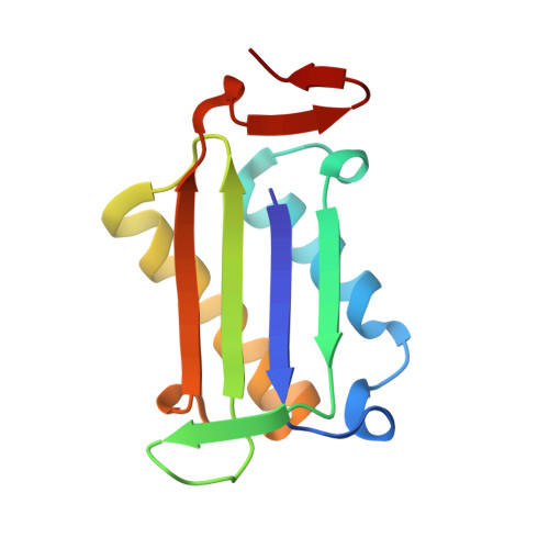
3SMB

Phenethylisothiocyanate Covalently Bound to Macrophage Migration Inhibitory Factor (MIF)


Experimental Data Snapshot

  • Method: X-RAY DIFFRACTION
  • Resolution: 1.60 Å
  • R-Value Free: 0.197 
  • R-Value Work: 0.181 

Starting Model: experimental
View more details

wwPDB Validation   3D Report Full Report


Ligand Structure Quality Assessment 


This is version 1.3 of the entry. See complete history


Literature

Structural interactions dictate the kinetics of macrophage migration inhibitory factor inhibition by different cancer-preventive isothiocyanates.

Crichlow, G.V.Fan, C.Keeler, C.Hodsdon, M.Lolis, E.J.

(2012) Biochemistry 51: 7506-7514

  • DOI: https://doi.org/10.1021/bi3005494
  • Primary Citation of Related Structures:  
    3SMB, 3SMC

  • PubMed Abstract: 

    Regulation of cellular processes by dietary nutrients is known to affect the likelihood of cancer development. One class of cancer-preventive nutrients, isothiocyanates (ITCs), derived from the consumption of cruciferous vegetables, is known to have various effects on cellular biochemistry. One target of ITCs is macrophage migration inhibitory factor (MIF), a widely expressed protein with known inflammatory, pro-tumorigenic, pro-angiogenic, and anti-apoptotic properties. MIF is covalently inhibited by a variety of ITCs, which in part may explain how they exert their cancer-preventive effects. We report the crystallographic structures of human MIF bound to phenethylisothiocyanate and to l-sulforaphane (dietary isothiocyanates derived from watercress and broccoli, respectively) and correlate structural features of these two isothiocyanates with their second-order rate constants for MIF inactivation. We also characterize changes in the MIF structure using nuclear magnetic resonance heteronuclear single-quantum coherence spectra of these complexes and observe many changes at the subunit interface. While a number of chemical shifts do not change, many of those that change do not have features similar in magnitude or direction for the two isothiocyanates. The difference in the binding modes of these two ITCs provides a means of using structure-activity relationships to reveal insights into MIF biological interactions. The results of this study provide a framework for the development of therapeutics that target MIF.


  • Organizational Affiliation

    Department of Pharmacology, Yale University School of Medicine, New Haven, CT 06510, USA.


Macromolecules
Find similar proteins by:  (by identity cutoff)  |  3D Structure
Entity ID: 1
MoleculeChains Sequence LengthOrganismDetailsImage
Macrophage migration inhibitory factor
A, B, C
114Homo sapiensMutation(s): 0 
Gene Names: GLIFMIFMMIF
EC: 5.3.2.1 (PDB Primary Data), 5.3.3.12 (PDB Primary Data)
UniProt & NIH Common Fund Data Resources
Find proteins for P14174 (Homo sapiens)
Explore P14174 
Go to UniProtKB:  P14174
PHAROS:  P14174
GTEx:  ENSG00000240972 
Entity Groups  
Sequence Clusters30% Identity50% Identity70% Identity90% Identity95% Identity100% Identity
UniProt GroupP14174
Sequence Annotations
Expand
  • Reference Sequence
Small Molecules
Ligands 3 Unique
IDChains Name / Formula / InChI Key2D Diagram3D Interactions
LE2
Query on LE2

Download Ideal Coordinates CCD File 
D [auth A],
J [auth B],
N [auth C]
N-(2-phenylethyl)thioformamide
C9 H11 N S
WVJCPUDEWCGDGL-UHFFFAOYSA-N
CL
Query on CL

Download Ideal Coordinates CCD File 
E [auth A]
F [auth A]
G [auth A]
H [auth A]
K [auth B]
E [auth A],
F [auth A],
G [auth A],
H [auth A],
K [auth B],
L [auth B],
O [auth C],
P [auth C],
Q [auth C]
CHLORIDE ION
Cl
VEXZGXHMUGYJMC-UHFFFAOYSA-M
NA
Query on NA

Download Ideal Coordinates CCD File 
I [auth A],
M [auth B],
R [auth C]
SODIUM ION
Na
FKNQFGJONOIPTF-UHFFFAOYSA-N
Experimental Data & Validation

Experimental Data

  • Method: X-RAY DIFFRACTION
  • Resolution: 1.60 Å
  • R-Value Free: 0.197 
  • R-Value Work: 0.181 
  • Space Group: P 21 21 21
Unit Cell:
Length ( Å )Angle ( ˚ )
a = 67.492α = 90
b = 67.67β = 90
c = 87.771γ = 90
Software Package:
Software NamePurpose
DENZOdata reduction
SCALEPACKdata scaling
AMoREphasing
CNSrefinement
PDB_EXTRACTdata extraction
CrystalCleardata collection
HKL-2000data reduction
HKL-2000data scaling

Structure Validation

View Full Validation Report


Ligand Structure Quality Assessment 


Entry History 

Deposition Data

Revision History  (Full details and data files)

  • Version 1.0: 2012-10-03
    Type: Initial release
  • Version 1.1: 2013-02-20
    Changes: Database references
  • Version 1.2: 2017-11-08
    Changes: Refinement description
  • Version 1.3: 2023-09-13
    Changes: Data collection, Database references, Derived calculations, Refinement description