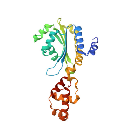
3SZT

Quorum Sensing Control Repressor, QscR, Bound to N-3-oxo-dodecanoyl-L-Homoserine Lactone


Experimental Data Snapshot

  • Method: X-RAY DIFFRACTION
  • Resolution: 2.55 Å
  • R-Value Free: 0.266 
  • R-Value Work: 0.223 
  • R-Value Observed: 0.225 

Starting Model: experimental
View more details

wwPDB Validation   3D Report Full Report


Ligand Structure Quality Assessment 


This is version 1.4 of the entry. See complete history


Literature

Crystal structure of QscR, a Pseudomonas aeruginosa quorum sensing signal receptor.

Lintz, M.J.Oinuma, K.Wysoczynski, C.L.Greenberg, E.P.Churchill, M.E.

(2011) Proc Natl Acad Sci U S A 108: 15763-15768

  • DOI: https://doi.org/10.1073/pnas.1112398108
  • Primary Citation of Related Structures:  
    3SZT

  • PubMed Abstract: 

    Acyl-homoserine lactone (AHL) quorum sensing controls gene expression in hundreds of Proteobacteria including a number of plant and animal pathogens. Generally, the AHL receptors are members of a family of related transcription factors, and although they have been targets for development of antivirulence therapeutics there is very little structural information about this class of bacterial receptors. We have determined the structure of the transcription factor, QscR, bound to N-3-oxo-dodecanoyl-homoserine lactone from the opportunistic human pathogen Pseudomonas aeruginosa at a resolution of 2.55 Å. The ligand-bound QscR is a dimer with a unique symmetric "cross-subunit" arrangement containing multiple dimerization interfaces involving both domains of each subunit. The QscR dimer appears poised to bind DNA. Predictions about signal binding and dimerization contacts were supported by studies of mutant QscR proteins in vivo. The acyl chain of the AHL is in close proximity to the dimerization interfaces. Our data are consistent with an allosteric mechanism of signal transmission in the regulation of DNA binding and thus virulence gene expression.


  • Organizational Affiliation

    Department of Pharmacology, Structural Biology and Biophysics Program, University of Colorado Denver School of Medicine, Aurora, CO 80045, USA.


Macromolecules
Find similar proteins by:  (by identity cutoff)  |  3D Structure
Entity ID: 1
MoleculeChains Sequence LengthOrganismDetailsImage
Quorum-sensing control repressor
A, B
237Pseudomonas aeruginosaMutation(s): 0 
Gene Names: PA1898qscR
UniProt
Find proteins for Q9RMS5 (Pseudomonas aeruginosa)
Explore Q9RMS5 
Go to UniProtKB:  Q9RMS5
Entity Groups  
Sequence Clusters30% Identity50% Identity70% Identity90% Identity95% Identity100% Identity
UniProt GroupQ9RMS5
Sequence Annotations
Expand
  • Reference Sequence
Small Molecules
Binding Affinity Annotations 
IDSourceBinding Affinity
OHN BindingDB:  3SZT EC50: min: 15, max: 22 (nM) from 2 assay(s)
Experimental Data & Validation

Experimental Data

  • Method: X-RAY DIFFRACTION
  • Resolution: 2.55 Å
  • R-Value Free: 0.266 
  • R-Value Work: 0.223 
  • R-Value Observed: 0.225 
  • Space Group: P 31 2 1
Unit Cell:
Length ( Å )Angle ( ˚ )
a = 94.862α = 90
b = 94.862β = 90
c = 104.953γ = 120
Software Package:
Software NamePurpose
Blu-Icedata collection
JBluIce-EPICSdata collection
PHASERphasing
REFMACrefinement
DENZOdata reduction
SCALEPACKdata scaling

Structure Validation

View Full Validation Report


Ligand Structure Quality Assessment 


Entry History 

Deposition Data

Revision History  (Full details and data files)

  • Version 1.0: 2011-09-28
    Type: Initial release
  • Version 1.1: 2011-10-05
    Changes: Database references
  • Version 1.2: 2017-11-08
    Changes: Refinement description
  • Version 1.3: 2024-02-28
    Changes: Data collection, Database references, Derived calculations
  • Version 1.4: 2024-04-03
    Changes: Refinement description