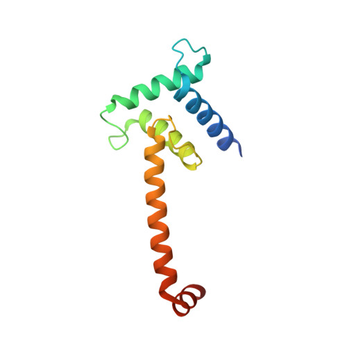
3T92

Crystal structure of the Taz2:C/EBPepsilon-TAD chimera protein


Experimental Data Snapshot

  • Method: X-RAY DIFFRACTION
  • Resolution: 1.50 Å
  • R-Value Free: 0.228 
  • R-Value Work: 0.178 
  • R-Value Observed: 0.180 

wwPDB Validation   3D Report Full Report


Ligand Structure Quality Assessment 


This is version 1.4 of the entry. See complete history


Literature

Structural insights into interactions of C/EBP transcriptional activators with the Taz2 domain of p300.

Bhaumik, P.Davis, J.Tropea, J.E.Cherry, S.Johnson, P.F.Miller, M.

(2014) Acta Crystallogr D Biol Crystallogr 70: 1914-1921

  • DOI: https://doi.org/10.1107/S1399004714009262
  • Primary Citation of Related Structures:  
    3T92

  • PubMed Abstract: 

    Members of the C/EBP family of transcription factors bind to the Taz2 domain of p300/CBP and mediate its phosphorylation through the recruitment of specific kinases. Short sequence motifs termed homology boxes A and B, which comprise their minimal transactivation domains (TADs), are conserved between C/EBP activators and are necessary for specific p300/CBP binding. A possible mode of interaction between C/EBP TADs and the p300 Taz2 domain was implied by the crystal structure of a chimeric protein composed of residues 1723-1818 of p300 Taz2 and residues 37-61 of C/EBPℇ. The segment corresponding to the C/EBPℇ TAD forms two orthogonally disposed helices connected by a short linker and interacts with the core structure of Taz2 from a symmetry-related molecule. It is proposed that other members of the C/EBP family interact with the Taz2 domain in the same manner. The position of the C/EBPℇ peptide on the Taz2 protein interaction surface suggests that the N-termini of C/EBP proteins are unbound in the C/EBP-p300 Taz2 complex. This observation is in agreement with the known location of the docking site of protein kinase HIPK2 in the C/EBPβ N-terminus, which associates with the C/EBPβ-p300 complex.


  • Organizational Affiliation

    Protein Structure Section, Macromolecular Crystallography Laboratory, National Cancer Institute at Frederick, Frederick, MD 21702, USA.


Macromolecules
Find similar proteins by:  (by identity cutoff)  |  3D Structure
Entity ID: 1
MoleculeChains Sequence LengthOrganismDetailsImage
HISTONE ACETYLTRANSFERASE P300 TAZ2-CCAAT/ENHANCER-BINDING PROTEIN EPSILON121Homo sapiensMutation(s): 4 
Gene Names: EP300P300
EC: 2.3.1.48 (PDB Primary Data), 2.3.1 (UniProt)
UniProt & NIH Common Fund Data Resources
Find proteins for Q15744 (Homo sapiens)
Explore Q15744 
Go to UniProtKB:  Q15744
PHAROS:  Q15744
GTEx:  ENSG00000092067 
Find proteins for Q09472 (Homo sapiens)
Explore Q09472 
Go to UniProtKB:  Q09472
PHAROS:  Q09472
GTEx:  ENSG00000100393 
Entity Groups  
Sequence Clusters30% Identity50% Identity70% Identity90% Identity95% Identity100% Identity
UniProt GroupsQ15744Q09472
Sequence Annotations
Expand
  • Reference Sequence
Small Molecules
Ligands 4 Unique
IDChains Name / Formula / InChI Key2D Diagram3D Interactions
TCE
Query on TCE

Download Ideal Coordinates CCD File 
E [auth A]3,3',3''-phosphanetriyltripropanoic acid
C9 H15 O6 P
PZBFGYYEXUXCOF-UHFFFAOYSA-N
TAM
Query on TAM

Download Ideal Coordinates CCD File 
F [auth A]TRIS(HYDROXYETHYL)AMINOMETHANE
C7 H17 N O3
GKODZWOPPOTFGA-UHFFFAOYSA-N
ZN
Query on ZN

Download Ideal Coordinates CCD File 
B [auth A],
C [auth A],
D [auth A]
ZINC ION
Zn
PTFCDOFLOPIGGS-UHFFFAOYSA-N
ACN
Query on ACN

Download Ideal Coordinates CCD File 
G [auth A]ACETONE
C3 H6 O
CSCPPACGZOOCGX-UHFFFAOYSA-N
Experimental Data & Validation

Experimental Data

  • Method: X-RAY DIFFRACTION
  • Resolution: 1.50 Å
  • R-Value Free: 0.228 
  • R-Value Work: 0.178 
  • R-Value Observed: 0.180 
  • Space Group: P 65
Unit Cell:
Length ( Å )Angle ( ˚ )
a = 47.86α = 90
b = 47.86β = 90
c = 104.13γ = 120
Software Package:
Software NamePurpose
MAR345dtbdata collection
MrBUMPphasing
REFMACrefinement
XDSdata reduction
XDSdata scaling

Structure Validation

View Full Validation Report


Ligand Structure Quality Assessment 


Entry History 

Deposition Data

Revision History  (Full details and data files)

  • Version 1.0: 2012-08-08
    Type: Initial release
  • Version 1.1: 2014-07-16
    Changes: Database references
  • Version 1.2: 2017-08-09
    Changes: Refinement description, Source and taxonomy
  • Version 1.3: 2018-10-10
    Changes: Data collection, Database references, Structure summary
  • Version 1.4: 2024-02-28
    Changes: Data collection, Database references, Derived calculations, Structure summary