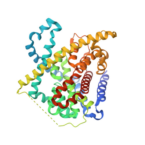
3TIJ

Crystal structure of a concentrative nucleoside transporter from Vibrio cholerae


Experimental Data Snapshot

  • Method: X-RAY DIFFRACTION
  • Resolution: 2.44 Å
  • R-Value Free: 0.228 
  • R-Value Work: 0.196 
  • R-Value Observed: 0.198 

wwPDB Validation   3D Report Full Report


Ligand Structure Quality Assessment 


This is version 1.2 of the entry. See complete history


Literature

Crystal structure of a concentrative nucleoside transporter from Vibrio cholerae at 2.4A

Johnson, Z.L.Cheong, C.G.Lee, S.Y.

(2012) Nature 483: 489-493

  • DOI: https://doi.org/10.1038/nature10882
  • Primary Citation of Related Structures:  
    3TIJ

  • PubMed Abstract: 

    Nucleosides are required for DNA and RNA synthesis, and the nucleoside adenosine has a function in a variety of signalling processes. Transport of nucleosides across cell membranes provides the major source of nucleosides in many cell types and is also responsible for the termination of adenosine signalling. As a result of their hydrophilic nature, nucleosides require a specialized class of integral membrane proteins, known as nucleoside transporters (NTs), for specific transport across cell membranes. In addition to nucleosides, NTs are important determinants for the transport of nucleoside-derived drugs across cell membranes. A wide range of nucleoside-derived drugs, including anticancer drugs (such as Ara-C and gemcitabine) and antiviral drugs (such as zidovudine and ribavirin), have been shown to depend, at least in part, on NTs for transport across cell membranes. Concentrative nucleoside transporters, members of the solute carrier transporter superfamily SLC28, use an ion gradient in the active transport of both nucleosides and nucleoside-derived drugs against their chemical gradients. The structural basis for selective ion-coupled nucleoside transport by concentrative nucleoside transporters is unknown. Here we present the crystal structure of a concentrative nucleoside transporter from Vibrio cholerae in complex with uridine at 2.4 Å. Our functional data show that, like its human orthologues, the transporter uses a sodium-ion gradient for nucleoside transport. The structure reveals the overall architecture of this class of transporter, unravels the molecular determinants for nucleoside and sodium binding, and provides a framework for understanding the mechanism of nucleoside and nucleoside drug transport across cell membranes.


  • Organizational Affiliation

    Department of Biochemistry and Ion Channel Research Unit, Duke University Medical Center, 2 Genome Court, Durham, North Carolina 27710, USA.


Macromolecules
Find similar proteins by:  (by identity cutoff)  |  3D Structure
Entity ID: 1
MoleculeChains Sequence LengthOrganismDetailsImage
NupC family protein424Vibrio choleraeMutation(s): 0 
Gene Names: VC_2352
Membrane Entity: Yes 
UniProt
Find proteins for Q9KPL5 (Vibrio cholerae serotype O1 (strain ATCC 39315 / El Tor Inaba N16961))
Explore Q9KPL5 
Go to UniProtKB:  Q9KPL5
Entity Groups  
Sequence Clusters30% Identity50% Identity70% Identity90% Identity95% Identity100% Identity
UniProt GroupQ9KPL5
Sequence Annotations
Expand
  • Reference Sequence
Small Molecules
Ligands 3 Unique
IDChains Name / Formula / InChI Key2D Diagram3D Interactions
DMU
Query on DMU

Download Ideal Coordinates CCD File 
C [auth A],
E [auth A],
F [auth A],
G [auth A],
H [auth A]
DECYL-BETA-D-MALTOPYRANOSIDE
C22 H42 O11
WOQQAWHSKSSAGF-WXFJLFHKSA-N
URI
Query on URI

Download Ideal Coordinates CCD File 
B [auth A]URIDINE
C9 H12 N2 O6
DRTQHJPVMGBUCF-XVFCMESISA-N
NA
Query on NA

Download Ideal Coordinates CCD File 
D [auth A]SODIUM ION
Na
FKNQFGJONOIPTF-UHFFFAOYSA-N
Experimental Data & Validation

Experimental Data

  • Method: X-RAY DIFFRACTION
  • Resolution: 2.44 Å
  • R-Value Free: 0.228 
  • R-Value Work: 0.196 
  • R-Value Observed: 0.198 
  • Space Group: P 63
Unit Cell:
Length ( Å )Angle ( ˚ )
a = 119.717α = 90
b = 119.717β = 90
c = 83.136γ = 120
Software Package:
Software NamePurpose
SERGUIdata collection
PHASERphasing
PHENIXrefinement
HKL-2000data reduction
HKL-2000data scaling

Structure Validation

View Full Validation Report


Ligand Structure Quality Assessment 


Entry History 

Deposition Data

Revision History  (Full details and data files)

  • Version 1.0: 2012-03-07
    Type: Initial release
  • Version 1.1: 2013-06-26
    Changes: Database references
  • Version 1.2: 2024-02-28
    Changes: Data collection, Database references, Derived calculations