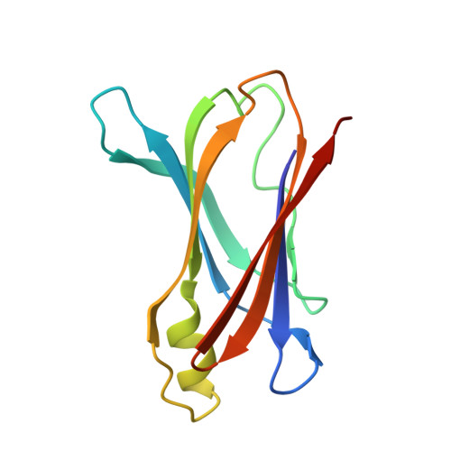
Hydrogen-bond network and pH sensitivity in transthyretin: Neutron crystal structure of human transthyretin
Yokoyama, T., Mizuguchi, M., Nabeshima, Y., Kusaka, K., Yamada, T., Hosoya, T., Ohhara, T., Kurihara, K., Tomoyori, K., Tanaka, I., Niimura, N.(2012) J Struct Biol 177: 283-290
- PubMed: 22248451
- DOI: https://doi.org/10.1016/j.jsb.2011.12.022
- Primary Citation of Related Structures:
3U2I, 3U2J - PubMed Abstract:
Transthyretin (TTR) is a tetrameric protein associated with human amyloidosis. In vitro, the formation of amyloid fibrils by TTR is known to be promoted by low pH. Here we show the neutron structure of TTR, focusing on the hydrogen bonds, protonation states and pH sensitivities. A large crystal was prepared at pD 7.4 for neutron protein crystallography. Neutron diffraction studies were conducted using the IBARAKI Biological Crystal Diffractometer with the time-of-flight method. The neutron structure solved at 2.0Å resolution revealed the protonation states of His88 and the detailed hydrogen-bond network depending on the protonation states of His88. This hydrogen-bond network is composed of Thr75, Trp79, His88, Ser112, Pro113, Thr118-B and four water molecules, and is involved in both monomer-monomer and dimer-dimer interactions, suggesting that the double protonation of His88 by acidification breaks the hydrogen-bond network and causes the destabilization of the TTR tetramer. In addition, the comparison with X-ray structure at pH 4.0 indicated that the protonation occurred to Asp74, His88 and Glu89 at pH 4.0. Our neutron model provides insights into the molecular stability of TTR related to the hydrogen-bond network, the pH sensitivity and the CH···O weak hydrogen bond.
Organizational Affiliation:
Frontier Research Center for Applied Atomic Sciences, Ibaraki University, 162-1 Shirakata, Tokai, Ibaraki 319-1106, Japan. [email protected]


