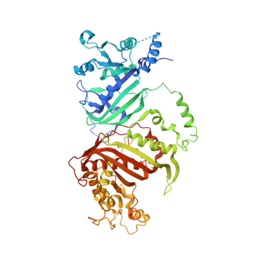
3UM6

Double mutant (A16V+S108T) Plasmodium falciparum DHFR-TS (T9/94) complexed with cycloguanil, NADPH and dUMP


Experimental Data Snapshot

  • Method: X-RAY DIFFRACTION
  • Resolution: 2.65 Å
  • R-Value Free: 0.257 
  • R-Value Work: 0.203 
  • R-Value Observed: 0.203 

wwPDB Validation   3D Report Full Report


Ligand Structure Quality Assessment 


This is version 1.2 of the entry. See complete history


Literature

Combined Spatial Limitation around Residues 16 and 108 of Plasmodium falciparum Dihydrofolate Reductase Explains Resistance to Cycloguanil.

Vanichtanankul, J.Taweechai, S.Uttamapinant, C.Chitnumsub, P.Vilaivan, T.Yuthavong, Y.Kamchonwongpaisan, S.

(2012) Antimicrob Agents Chemother 56: 3928-3935

  • DOI: https://doi.org/10.1128/AAC.00301-12
  • Primary Citation of Related Structures:  
    3UM5, 3UM6, 3UM8

  • PubMed Abstract: 

    Natural mutations of Plasmodium falciparum dihydrofolate reductase (PfDHFR) at A16V and S108T specifically confer resistance to cycloguanil (CYC) but not to pyrimethamine (PYR). In order to understand the nature of CYC resistance, the effects of various mutations at A16 on substrate and inhibitor binding were examined. Three series of mutations at A16 with or without the S108T/N mutation were generated. Only three mutants with small side chains at residue 16 (G, C, and S) were viable from bacterial complementation assay in the S108 series, whereas these three and an additional four mutants (T, V, M, and I) with slightly larger side chains were viable with simultaneous S108T mutation. Among these combinations, the A16V+S108T mutant was the most CYC resistant, and all of the S108T series ranged from being highly to moderately sensitive to PYR. In the S108N series, a strict requirement for alanine was observed at position 16. Crystal structure analyses reveal that in PfDHFR-TS variant T9/94 (A16V+S108T) complexed with CYC, the ligand has substantial steric conflicts with the side chains of both A16V and S108T, whereas in the complex with PYR, the ligand only showed mild conflict with S108T. CYC analogs designed to avoid such conflicts improved the binding affinity of the mutant enzymes. These results show that there is greater spatial limitation around the S108T/N residue when combined with the limitation imposed by A16V. The limitation of mutation of this series provides opportunities for drug design and development against antifolate-resistant malaria.


  • Organizational Affiliation

    National Centre for Genetic Engineering and Biotechnology, National Science and Technology Development Agency, Klong Luang, Pathumthani, Thailand.


Macromolecules
Find similar proteins by:  (by identity cutoff)  |  3D Structure
Entity ID: 1
MoleculeChains Sequence LengthOrganismDetailsImage
Bifunctional dihydrofolate reductase-thymidylate synthase
A, B
608Plasmodium falciparumMutation(s): 2 
Gene Names: dhfr-ts
EC: 1.5.1.3 (PDB Primary Data), 2.1.1.45 (PDB Primary Data)
UniProt
Find proteins for A7UD81 (Plasmodium falciparum)
Explore A7UD81 
Go to UniProtKB:  A7UD81
Entity Groups  
Sequence Clusters30% Identity50% Identity70% Identity90% Identity95% Identity100% Identity
UniProt GroupA7UD81
Sequence Annotations
Expand
  • Reference Sequence
Small Molecules
Ligands 3 Unique
IDChains Name / Formula / InChI Key2D Diagram3D Interactions
NDP
Query on NDP

Download Ideal Coordinates CCD File 
D [auth A],
G [auth B]
NADPH DIHYDRO-NICOTINAMIDE-ADENINE-DINUCLEOTIDE PHOSPHATE
C21 H30 N7 O17 P3
ACFIXJIJDZMPPO-NNYOXOHSSA-N
UMP
Query on UMP

Download Ideal Coordinates CCD File 
E [auth A],
H [auth B]
2'-DEOXYURIDINE 5'-MONOPHOSPHATE
C9 H13 N2 O8 P
JSRLJPSBLDHEIO-SHYZEUOFSA-N
1CY
Query on 1CY

Download Ideal Coordinates CCD File 
C [auth A],
F [auth B]
1-(4-chlorophenyl)-6,6-dimethyl-1,6-dihydro-1,3,5-triazine-2,4-diamine
C11 H14 Cl N5
QMNFFXRFOJIOKZ-UHFFFAOYSA-N
Binding Affinity Annotations 
IDSourceBinding Affinity
1CY BindingDB:  3UM6 Ki: min: 0.3, max: 2130 (nM) from 17 assay(s)
IC50: min: 1.5, max: 5800 (nM) from 6 assay(s)
Experimental Data & Validation

Experimental Data

  • Method: X-RAY DIFFRACTION
  • Resolution: 2.65 Å
  • R-Value Free: 0.257 
  • R-Value Work: 0.203 
  • R-Value Observed: 0.203 
  • Space Group: P 21 21 21
Unit Cell:
Length ( Å )Angle ( ˚ )
a = 56.696α = 90
b = 155.549β = 90
c = 164.962γ = 90
Software Package:
Software NamePurpose
SCALEPACKdata scaling
AMoREphasing
CNSrefinement
PDB_EXTRACTdata extraction
DENZOdata reduction

Structure Validation

View Full Validation Report


Ligand Structure Quality Assessment 


Entry History 

Deposition Data

Revision History  (Full details and data files)

  • Version 1.0: 2012-07-18
    Type: Initial release
  • Version 1.1: 2017-11-08
    Changes: Refinement description
  • Version 1.2: 2024-03-20
    Changes: Data collection, Database references, Derived calculations