3WFT

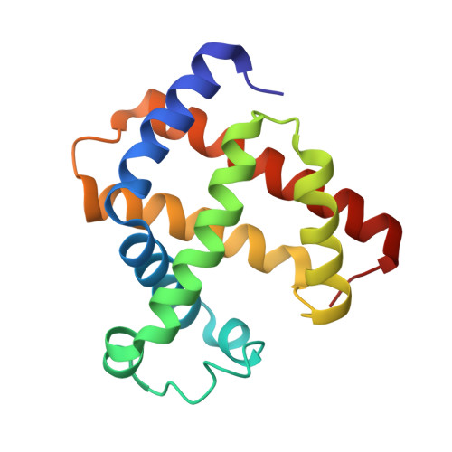
Crystal structure of horse heart myoglobin reconstituted with cobalt(II) tetradehydrocorrin


Experimental Data Snapshot

  • Method: X-RAY DIFFRACTION
  • Resolution: 1.30 Å
  • R-Value Free: 0.183 
  • R-Value Work: 0.126 
  • R-Value Observed: 0.129 

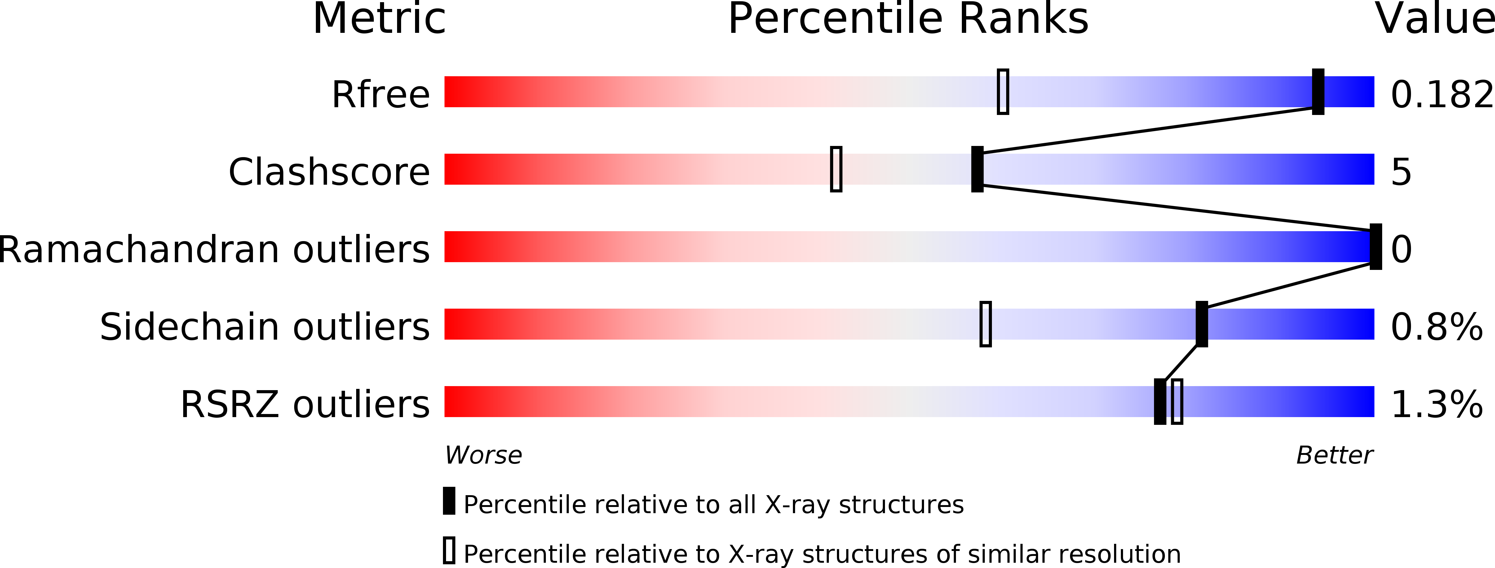
wwPDB Validation   3D Report Full Report


Ligand Structure Quality Assessment 


This is version 1.2 of the entry. See complete history


Literature

Co(II)/Co(I) reduction-induced axial histidine-flipping in myoglobin reconstituted with a cobalt tetradehydrocorrin as a methionine synthase model.

Hayashi, T.Morita, Y.Mizohata, E.Oohora, K.Ohbayashi, J.Inoue, T.Hisaeda, Y.

(2014) Chem Commun (Camb) 50: 12560-12563

  • DOI: https://doi.org/10.1039/c4cc05448b
  • Primary Citation of Related Structures:  
    3WFT, 3WFU

  • PubMed Abstract: 

    A conjugate between apomyoglobin and cobalt tetradehydrocorrin was prepared to replicate the coordination behavior of cob(I)alamin in methionine synthase. X-ray crystallography reveals that the tetra-coordinated Co(I) species is formed through the cleavage of the axial Co-His93 ligation after the reduction of the penta-coordinated Co(II) cofactor in the heme pocket.


  • Organizational Affiliation

    Department of Applied Chemistry, Graduate School of Engineering, Osaka University, Suita 565-0871, Japan. [email protected].


Macromolecules
Find similar proteins by:  (by identity cutoff)  |  3D Structure
Entity ID: 1
MoleculeChains Sequence LengthOrganismDetailsImage
Myoglobin153Equus caballusMutation(s): 0 
EC: 1.11.1 (UniProt), 1.7 (UniProt)
UniProt
Find proteins for P68082 (Equus caballus)
Explore P68082 
Go to UniProtKB:  P68082
Entity Groups  
Sequence Clusters30% Identity50% Identity70% Identity90% Identity95% Identity100% Identity
UniProt GroupP68082
Sequence Annotations
Expand
  • Reference Sequence
Small Molecules
Ligands 4 Unique
IDChains Name / Formula / InChI Key2D Diagram3D Interactions
J1S
Query on J1S

Download Ideal Coordinates CCD File 
C [auth A](1S,19S) cobalt tetradehydrocorrin
C33 H37 Co N4 O4
DBXMOQHUTDEXAY-MLGYITDRSA-M
J1R
Query on J1R

Download Ideal Coordinates CCD File 
B [auth A](1R,19R) cobalt tetradehydrocorrin
C33 H37 Co N4 O4
DBXMOQHUTDEXAY-ZNBLAKOKSA-M
SO4
Query on SO4

Download Ideal Coordinates CCD File 
D [auth A],
E [auth A]
SULFATE ION
O4 S
QAOWNCQODCNURD-UHFFFAOYSA-L
GOL
Query on GOL

Download Ideal Coordinates CCD File 
F [auth A],
G [auth A],
H [auth A]
GLYCEROL
C3 H8 O3
PEDCQBHIVMGVHV-UHFFFAOYSA-N
Experimental Data & Validation

Experimental Data

  • Method: X-RAY DIFFRACTION
  • Resolution: 1.30 Å
  • R-Value Free: 0.183 
  • R-Value Work: 0.126 
  • R-Value Observed: 0.129 
  • Space Group: P 1 21 1
Unit Cell:
Length ( Å )Angle ( ˚ )
a = 34.975α = 90
b = 28.987β = 106.14
c = 63.214γ = 90
Software Package:
Software NamePurpose
SERGUIdata collection
MOLREPphasing
REFMACrefinement
HKL-2000data reduction
HKL-2000data scaling

Structure Validation

View Full Validation Report


Ligand Structure Quality Assessment 


Entry History 

Deposition Data

Revision History  (Full details and data files)

  • Version 1.0: 2014-12-03
    Type: Initial release
  • Version 1.1: 2017-11-22
    Changes: Refinement description
  • Version 1.2: 2024-03-20
    Changes: Data collection, Database references, Derived calculations