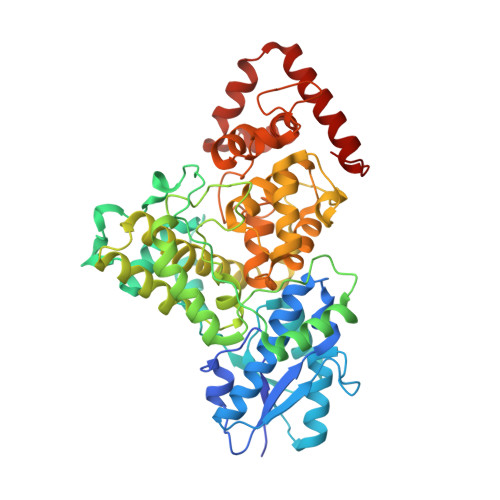
3ZXS

Cryptochrome B from Rhodobacter sphaeroides


Experimental Data Snapshot

  • Method: X-RAY DIFFRACTION
  • Resolution: 2.70 Å
  • R-Value Free: 0.228 
  • R-Value Work: 0.198 
  • R-Value Observed: 0.199 

wwPDB Validation   3D Report Full Report


Ligand Structure Quality Assessment 


This is version 1.3 of the entry. See complete history


Literature

Cryb from Rhodobacter Sphaeroides: A Unique Class of Cryptochromes with New Cofactors.

Geisselbrecht, Y.Fruhwirth, S.Schroeder, C.Pierik, A.J.Klug, G.Essen, L.-O.

(2012) EMBO Rep 13: 223

  • DOI: https://doi.org/10.1038/embor.2012.2
  • Primary Citation of Related Structures:  
    3ZXS

  • PubMed Abstract: 

    Cryptochromes and photolyases are structurally related but have different biological functions in signalling and DNA repair. Proteobacteria and cyanobacteria harbour a new class of cryptochromes, called CryPro. We have solved the 2.7 Å structure of one of its members, cryptochrome B from Rhodobacter sphaeroides, which is a regulator of photosynthesis gene expression. The structure reveals that, in addition to the photolyase-like fold, CryB contains two cofactors only conserved in the CryPro subfamily: 6,7-dimethyl-8-ribityl-lumazine in the antenna-binding domain and a [4Fe-4S] cluster within the catalytic domain. The latter closely resembles the iron-sulphur cluster harbouring the large primase subunit PriL, indicating that PriL is evolutionarily related to the CryPro class of cryptochromes.


  • Organizational Affiliation

    Department of Chemistry-Institute of Biochemistry, Philipps-University, Marburg, Germany.


Macromolecules
Find similar proteins by:  (by identity cutoff)  |  3D Structure
Entity ID: 1
MoleculeChains Sequence LengthOrganismDetailsImage
CRYPTOCHROME B
A, B, C
522Cereibacter sphaeroides 2.4.1Mutation(s): 0 
UniProt
Find proteins for Q3IXP1 (Cereibacter sphaeroides (strain ATCC 17023 / DSM 158 / JCM 6121 / CCUG 31486 / LMG 2827 / NBRC 12203 / NCIMB 8253 / ATH 2.4.1.))
Explore Q3IXP1 
Go to UniProtKB:  Q3IXP1
Entity Groups  
Sequence Clusters30% Identity50% Identity70% Identity90% Identity95% Identity100% Identity
UniProt GroupQ3IXP1
Sequence Annotations
Expand
  • Reference Sequence
Small Molecules
Ligands 5 Unique
IDChains Name / Formula / InChI Key2D Diagram3D Interactions
FAD
Query on FAD

Download Ideal Coordinates CCD File 
D [auth A],
I [auth B],
N [auth C]
FLAVIN-ADENINE DINUCLEOTIDE
C27 H33 N9 O15 P2
VWWQXMAJTJZDQX-UYBVJOGSSA-N
SF4
Query on SF4

Download Ideal Coordinates CCD File 
E [auth A],
J [auth B],
O [auth C]
IRON/SULFUR CLUSTER
Fe4 S4
LJBDFODJNLIPKO-UHFFFAOYSA-N
DLZ
Query on DLZ

Download Ideal Coordinates CCD File 
F [auth A],
K [auth B],
P [auth C]
1-deoxy-1-(6,7-dimethyl-2,4-dioxo-3,4-dihydropteridin-8(2H)-yl)-D-ribitol
C13 H18 N4 O6
SXDXRJZUAJBNFL-XKSSXDPKSA-N
GD
Query on GD

Download Ideal Coordinates CCD File 
H [auth A],
M [auth B],
R [auth C]
GADOLINIUM ATOM
Gd
UIWYJDYFSGRHKR-UHFFFAOYSA-N
MG
Query on MG

Download Ideal Coordinates CCD File 
G [auth A],
L [auth B],
Q [auth C]
MAGNESIUM ION
Mg
JLVVSXFLKOJNIY-UHFFFAOYSA-N
Experimental Data & Validation

Experimental Data

  • Method: X-RAY DIFFRACTION
  • Resolution: 2.70 Å
  • R-Value Free: 0.228 
  • R-Value Work: 0.198 
  • R-Value Observed: 0.199 
  • Space Group: P 65 2 2
Unit Cell:
Length ( Å )Angle ( ˚ )
a = 137.84α = 90
b = 137.84β = 90
c = 521.94γ = 120
Software Package:
Software NamePurpose
REFMACrefinement
XDSdata reduction
XSCALEdata scaling
PHENIXphasing

Structure Validation

View Full Validation Report


Ligand Structure Quality Assessment 


Entry History 

Deposition Data

Revision History  (Full details and data files)

  • Version 1.0: 2012-02-15
    Type: Initial release
  • Version 1.1: 2012-03-28
    Changes: Other
  • Version 1.2: 2014-08-13
    Changes: Refinement description
  • Version 1.3: 2024-05-08
    Changes: Data collection, Database references, Derived calculations, Other, Refinement description