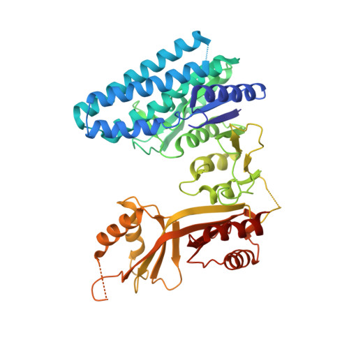
3C1N

Crystal Structure of Allosteric Inhibition Threonine-sensitive Aspartokinase from Methanococcus jannaschii with L-threonine


Experimental Data Snapshot

  • Method: X-RAY DIFFRACTION
  • Resolution: 2.72 Å
  • R-Value Free: 0.290 
  • R-Value Work: 0.236 
  • R-Value Observed: 0.239 

Starting Model: experimental
View more details

wwPDB Validation   3D Report Full Report


This is version 1.3 of the entry. See complete history


Literature

The Structural Basis for Allosteric Inhibition of a Threonine-sensitive Aspartokinase.

Liu, X.Pavlovsky, A.G.Viola, R.E.

(2008) J Biol Chem 283: 16216-16225

  • DOI: https://doi.org/10.1074/jbc.M800760200
  • Primary Citation of Related Structures:  
    3C1M, 3C1N, 3C20

  • PubMed Abstract: 

    The commitment step to the aspartate pathway of amino acid biosynthesis is the phosphorylation of aspartic acid catalyzed by aspartokinase (AK). Most microorganisms and plants have multiple forms of this enzyme, and many of these isofunctional enzymes are subject to feedback regulation by the end products of the pathway. However, the archeal species Methanococcus jannaschii has only a single, monofunctional form of AK. The substrate l-aspartate binds to this recombinant enzyme in two different orientations, providing the first structural evidence supporting the relaxed regiospecificity previously observed with several alternative substrates of Escherichia coli AK ( Angeles, T. S., Hunsley, J. R., and Viola, R. E. (1992) Biochemistry 31, 799-805 ). Binding of the nucleotide substrate triggers significant domain movements that result in a more compact quaternary structure. In contrast, the highly cooperative binding of the allosteric regulator l-threonine to multiple sites on this dimer of dimers leads to an open enzyme structure. A comparison of these structures supports a mechanism for allosteric regulation in which the domain movements induced by threonine binding causes displacement of the substrates from the enzyme, resulting in a relaxed, inactive conformation.


  • Organizational Affiliation

    Department of Chemistry, University of Toledo, Toledo, Ohio 43606.


Macromolecules
Find similar proteins by:  (by identity cutoff)  |  3D Structure
Entity ID: 1
MoleculeChains Sequence LengthOrganismDetailsImage
Probable aspartokinase
A, B, C, D
473Methanocaldococcus jannaschiiMutation(s): 0 
Gene Names: MJ0571
EC: 2.7.2.4
UniProt
Find proteins for Q57991 (Methanocaldococcus jannaschii (strain ATCC 43067 / DSM 2661 / JAL-1 / JCM 10045 / NBRC 100440))
Explore Q57991 
Go to UniProtKB:  Q57991
Entity Groups  
Sequence Clusters30% Identity50% Identity70% Identity90% Identity95% Identity100% Identity
UniProt GroupQ57991
Sequence Annotations
Expand
  • Reference Sequence
Experimental Data & Validation

Experimental Data

  • Method: X-RAY DIFFRACTION
  • Resolution: 2.72 Å
  • R-Value Free: 0.290 
  • R-Value Work: 0.236 
  • R-Value Observed: 0.239 
  • Space Group: P 21 21 21
Unit Cell:
Length ( Å )Angle ( ˚ )
a = 110.039α = 90
b = 144.317β = 90
c = 155.358γ = 90
Software Package:
Software NamePurpose
DENZOdata reduction
SCALEPACKdata scaling
PHASERphasing
REFMACrefinement
PDB_EXTRACTdata extraction
SERGUIdata collection

Structure Validation

View Full Validation Report


Entry History 

Deposition Data

Revision History  (Full details and data files)

  • Version 1.0: 2008-03-25
    Type: Initial release
  • Version 1.1: 2011-07-13
    Changes: Advisory, Version format compliance
  • Version 1.2: 2017-10-25
    Changes: Refinement description
  • Version 1.3: 2023-08-30
    Changes: Data collection, Database references, Derived calculations, Refinement description