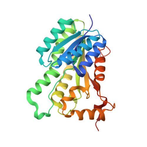
3OID

Crystal Structure of Enoyl-ACP Reductases III (FabL) from B. subtilis (complex with NADP and TCL)


Experimental Data Snapshot

  • Method: X-RAY DIFFRACTION
  • Resolution: 1.80 Å
  • R-Value Free: 0.181 
  • R-Value Work: 0.149 
  • R-Value Observed: 0.151 

Starting Model: experimental
View more details

wwPDB Validation   3D Report Full Report


Ligand Structure Quality Assessment 


This is version 1.2 of the entry. See complete history


Literature

Crystal Structures of Enoyl-ACP Reductases I (FabI) and III (FabL) from B. subtilis

Kim, K.-H.Ha, B.H.Kim, S.J.Hong, S.K.Hwang, K.Y.Kim, E.E.

(2011) J Mol Biol 406: 403-415

  • DOI: https://doi.org/10.1016/j.jmb.2010.12.003
  • Primary Citation of Related Structures:  
    3OIC, 3OID, 3OIF, 3OIG

  • PubMed Abstract: 

    Enoyl-[acyl carrier protein] (ACP) reductase (ENR) is a key enzyme in type II fatty acid synthesis that catalyzes the last step in each elongation cycle. Therefore, it has been considered as a target for antibiotics. However, recent studies indicate that some pathogens have more than one ENR; in particular, Bacillus subtilis has two ENRs, FabI and FabL. The crystal structures of the ternary complexes of BsFaBI and BsFabL are found as a homotetramer showing the same overall structure despite a sequence identity of only 24%. The positions of the catalytic dyad of Tyr-(Xaa)(6)-Lys in FabL are almost identical to that of FabI, but a detailed structural analysis shows that FabL shares more structural similarities with FabG and other members of the SDR (short-chain alcohol dehydrogenase/reductase) family. The apo FabL structure shows significantly different conformations at the cofactor and the substrate-binding regions, and this resulted in a totally different tetrameric arrangement reflecting the flexibility of these regions in the absence of the cofactor and substrate/inhibitor.


  • Organizational Affiliation

    Life Sciences Division, Korea Institute of Science and Technology, 39-1 Hawolkok-dong, Sungbuk-gu, Seoul 136-791, South Korea.


Macromolecules
Find similar proteins by:  (by identity cutoff)  |  3D Structure
Entity ID: 1
MoleculeChains Sequence LengthOrganismDetailsImage
Enoyl-[acyl-carrier-protein] reductase [NADPH]
A, B, C, D
258Bacillus subtilisMutation(s): 0 
Gene Names: FabL
EC: 1.3.1.10 (PDB Primary Data), 1.3.1.104 (UniProt)
UniProt
Find proteins for P71079 (Bacillus subtilis (strain 168))
Explore P71079 
Go to UniProtKB:  P71079
Entity Groups  
Sequence Clusters30% Identity50% Identity70% Identity90% Identity95% Identity100% Identity
UniProt GroupP71079
Sequence Annotations
Expand
  • Reference Sequence
Small Molecules
Ligands 2 Unique
IDChains Name / Formula / InChI Key2D Diagram3D Interactions
NDP
Query on NDP

Download Ideal Coordinates CCD File 
F [auth A],
H [auth B],
J [auth C],
L [auth D]
NADPH DIHYDRO-NICOTINAMIDE-ADENINE-DINUCLEOTIDE PHOSPHATE
C21 H30 N7 O17 P3
ACFIXJIJDZMPPO-NNYOXOHSSA-N
TCL
Query on TCL

Download Ideal Coordinates CCD File 
E [auth A],
G [auth B],
I [auth C],
K [auth D]
TRICLOSAN
C12 H7 Cl3 O2
XEFQLINVKFYRCS-UHFFFAOYSA-N
Experimental Data & Validation

Experimental Data

  • Method: X-RAY DIFFRACTION
  • Resolution: 1.80 Å
  • R-Value Free: 0.181 
  • R-Value Work: 0.149 
  • R-Value Observed: 0.151 
  • Space Group: P 1 21 1
Unit Cell:
Length ( Å )Angle ( ˚ )
a = 58.376α = 90
b = 110.683β = 91.82
c = 85.169γ = 90
Software Package:
Software NamePurpose
ADSCdata collection
CNSrefinement
REFMACrefinement
HKL-2000data reduction
HKL-2000data scaling
CNSphasing

Structure Validation

View Full Validation Report


Ligand Structure Quality Assessment 


Entry History 

Deposition Data

Revision History  (Full details and data files)

  • Version 1.0: 2011-01-05
    Type: Initial release
  • Version 1.1: 2011-07-13
    Changes: Version format compliance
  • Version 1.2: 2023-11-01
    Changes: Data collection, Database references, Derived calculations, Refinement description