3W67

Crystal structure of mouse alpha-tocopherol transfer protein in complex with alpha-tocopherol and phosphatidylinositol-(3,4)-bisphosphate


Experimental Data Snapshot

  • Method: X-RAY DIFFRACTION
  • Resolution: 2.61 Å
  • R-Value Free: 0.260 
  • R-Value Work: 0.211 
  • R-Value Observed: 0.213 

Starting Model: experimental
View more details

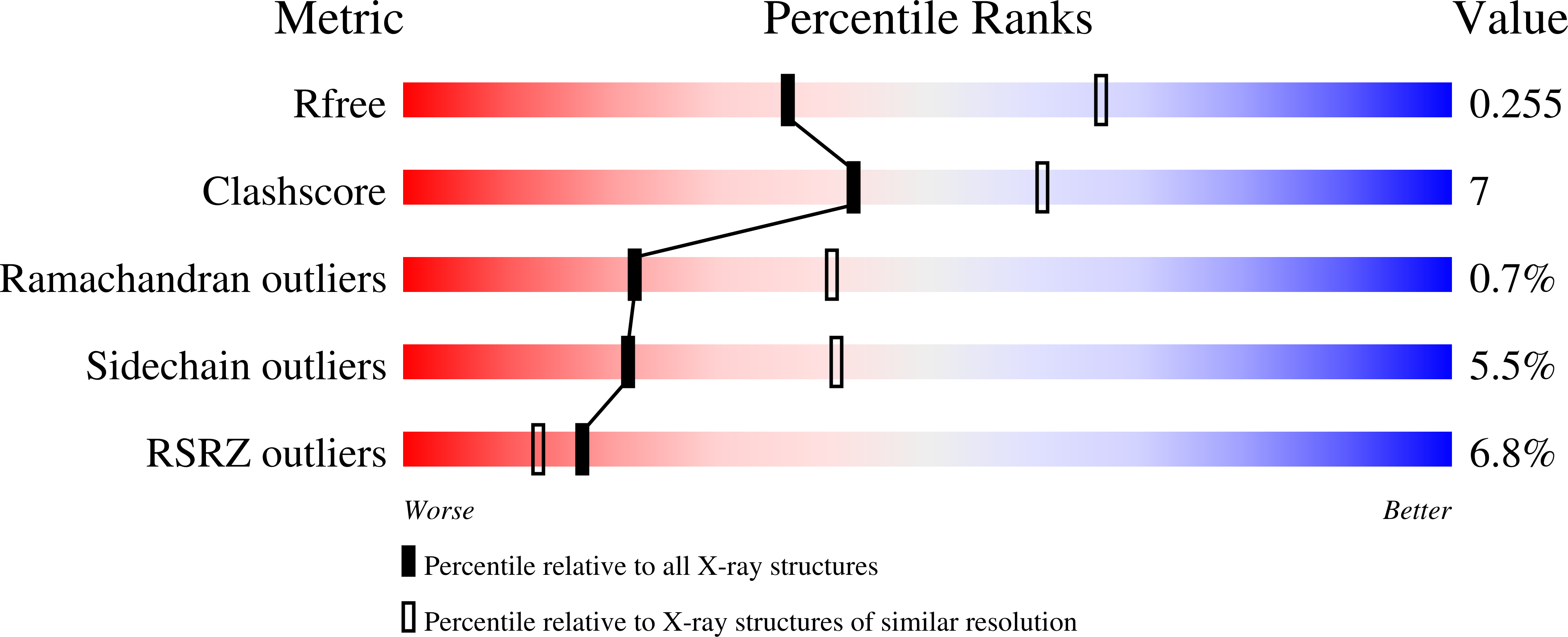
wwPDB Validation   3D Report Full Report


Ligand Structure Quality Assessment 


This is version 1.2 of the entry. See complete history


Literature

Impaired alpha-TTP-PIPs interaction underlies familial vitamin E deficiency

Kono, N.Ohto, U.Hiramatsu, T.Urabe, M.Uchida, Y.Satow, Y.Arai, H.

(2013) Science 340: 1106-1110

  • DOI: https://doi.org/10.1126/science.1233508
  • Primary Citation of Related Structures:  
    3W67, 3W68

  • PubMed Abstract: 

    α-Tocopherol (vitamin E) transfer protein (α-TTP) regulates the secretion of α-tocopherol from liver cells. Missense mutations of some arginine residues at the surface of α-TTP cause severe vitamin E deficiency in humans, but the role of these residues is unclear. Here, we found that wild-type α-TTP bound phosphatidylinositol phosphates (PIPs), whereas the arginine mutants did not. In addition, PIPs in the target membrane promoted the intermembrane transfer of α-tocopherol by α-TTP. The crystal structure of the α-TTP-PIPs complex revealed that the disease-related arginine residues interacted with phosphate groups of the PIPs and that the PIPs binding caused the lid of the α-tocopherol-binding pocket to open. Thus, PIPs have a role in promoting the release of a ligand from a lipid-transfer protein.


  • Organizational Affiliation

    Graduate School of Pharmaceutical Sciences, University of Tokyo, Tokyo, Japan.


Macromolecules
Find similar proteins by:  (by identity cutoff)  |  3D Structure
Entity ID: 1
MoleculeChains Sequence LengthOrganismDetailsImage
Alpha-tocopherol transfer protein
A, B, C, D
266Mus musculusMutation(s): 0 
Gene Names: Ttpa
UniProt
Find proteins for Q8BWP5 (Mus musculus)
Explore Q8BWP5 
Go to UniProtKB:  Q8BWP5
Entity Groups  
Sequence Clusters30% Identity50% Identity70% Identity90% Identity95% Identity100% Identity
UniProt GroupQ8BWP5
Sequence Annotations
Expand
  • Reference Sequence
Small Molecules
Ligands 2 Unique
IDChains Name / Formula / InChI Key2D Diagram3D Interactions
3PT
Query on 3PT

Download Ideal Coordinates CCD File 
F [auth A],
H [auth B],
J [auth C],
L [auth D]
(2R)-3-{[(S)-hydroxy{[(1S,2R,3R,4S,5S,6S)-2,3,6-trihydroxy-4,5-bis(phosphonooxy)cyclohexyl]oxy}phosphoryl]oxy}propane-1,2-diyl dibutanoate
C17 H33 O19 P3
DHAFWWKSHUBGAH-DXOPXGNRSA-N
VIV
Query on VIV

Download Ideal Coordinates CCD File 
E [auth A],
G [auth B],
I [auth C],
K [auth D]
(2R)-2,5,7,8-TETRAMETHYL-2-[(4R,8R)-4,8,12-TRIMETHYLTRIDECYL]CHROMAN-6-OL
C29 H50 O2
GVJHHUAWPYXKBD-IEOSBIPESA-N
Binding Affinity Annotations 
IDSourceBinding Affinity
VIV BindingDB:  3W67 Kd: 2420 (nM) from 1 assay(s)
Experimental Data & Validation

Experimental Data

  • Method: X-RAY DIFFRACTION
  • Resolution: 2.61 Å
  • R-Value Free: 0.260 
  • R-Value Work: 0.211 
  • R-Value Observed: 0.213 
  • Space Group: P 1
Unit Cell:
Length ( Å )Angle ( ˚ )
a = 58.285α = 100.24
b = 69.665β = 109.72
c = 87.183γ = 100.46
Software Package:
Software NamePurpose
MOLREPphasing
REFMACrefinement
HKL-2000data reduction
HKL-2000data scaling

Structure Validation

View Full Validation Report


Ligand Structure Quality Assessment 


Entry History 

Deposition Data

Revision History  (Full details and data files)

  • Version 1.0: 2013-05-01
    Type: Initial release
  • Version 1.1: 2013-09-04
    Changes: Database references
  • Version 1.2: 2023-11-08
    Changes: Data collection, Database references, Derived calculations, Refinement description